. 2021:1:1.
doi: 10.25259/ajbps_6_2021. Epub 2021 Nov 1.

Selenium-Dependent Read Through of the Conserved 3'-Terminal UGA Stop Codon of HIV-1 nef

Affiliations

Selenium-Dependent Read Through of the Conserved 3'-Terminal UGA Stop Codon of HIV-1 nef

Lakmini S Premadasa et al. Am J Biopharm Pharm Sci. 2021.

Abstract

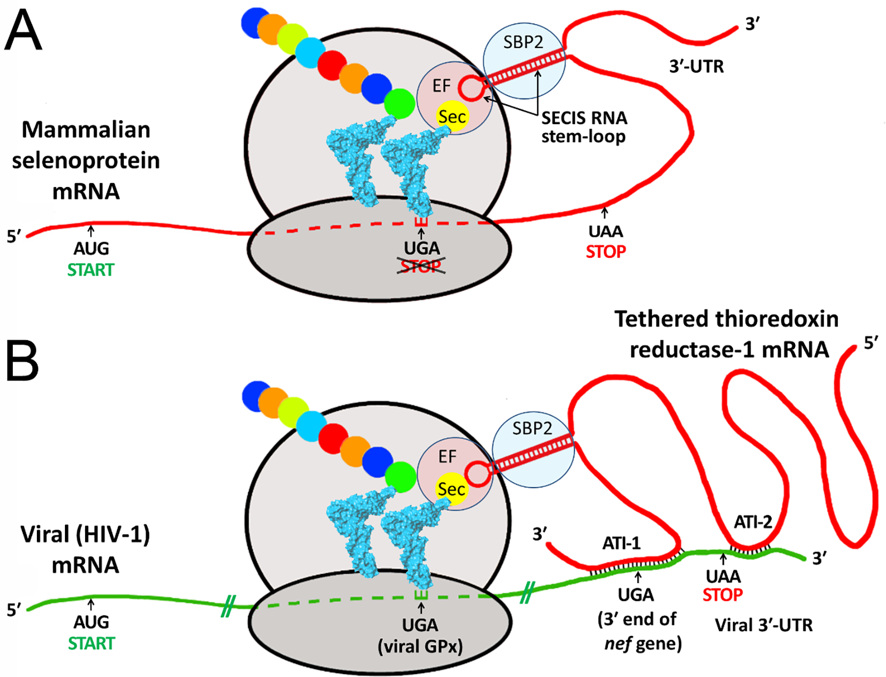
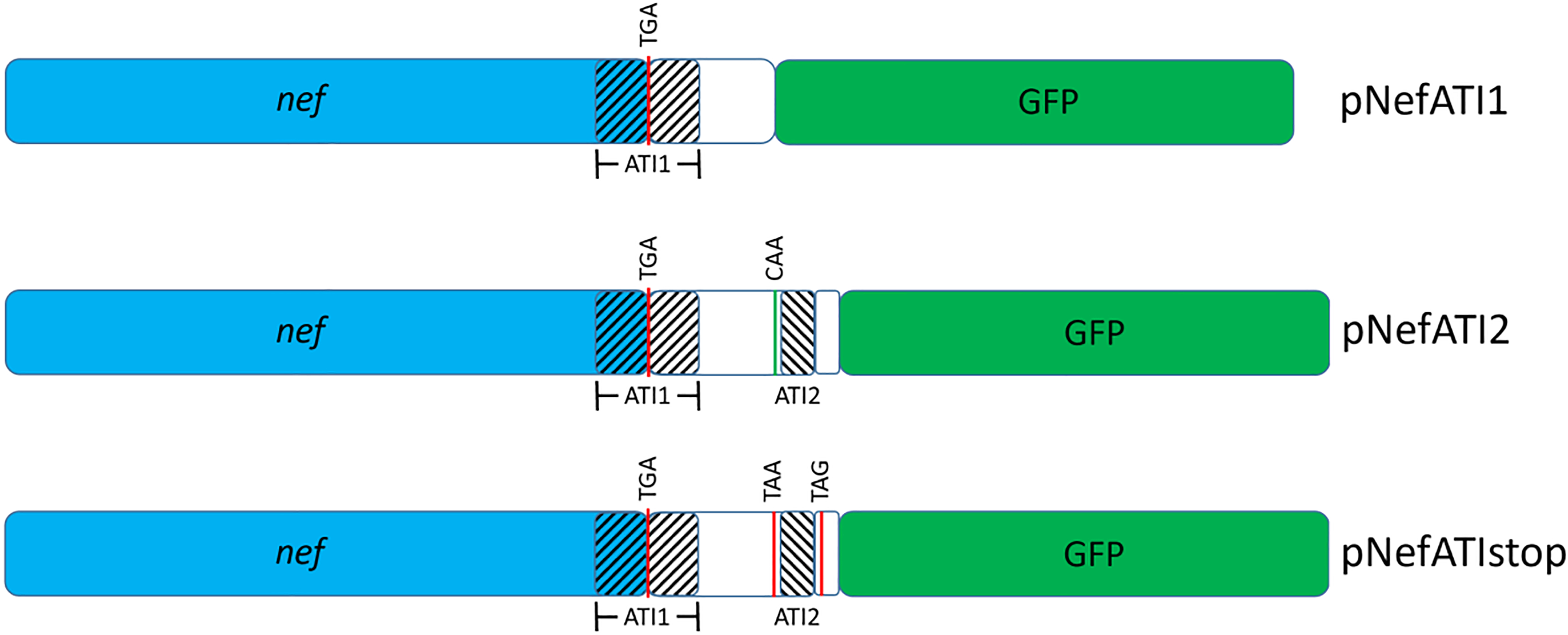
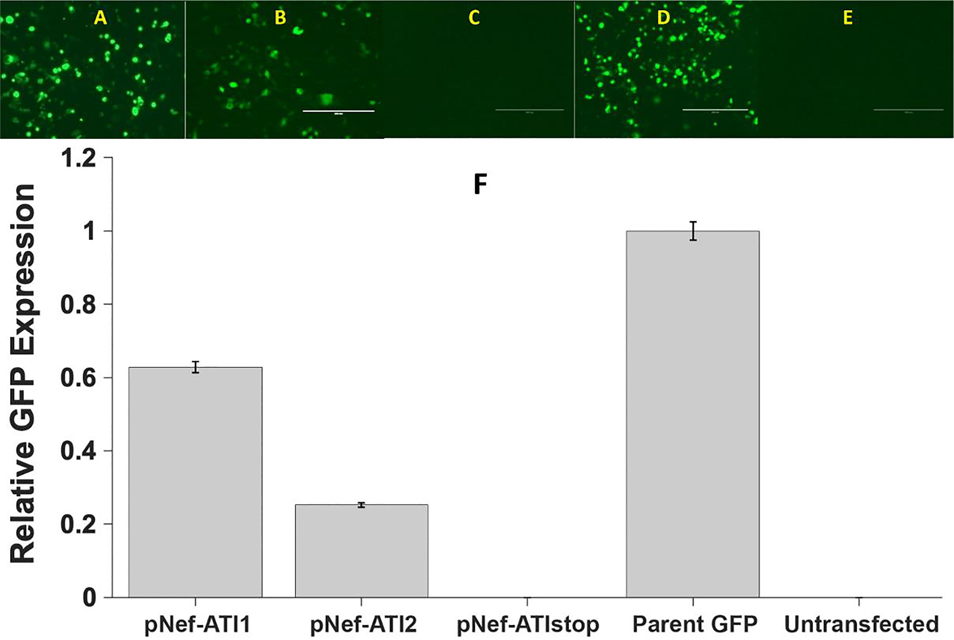
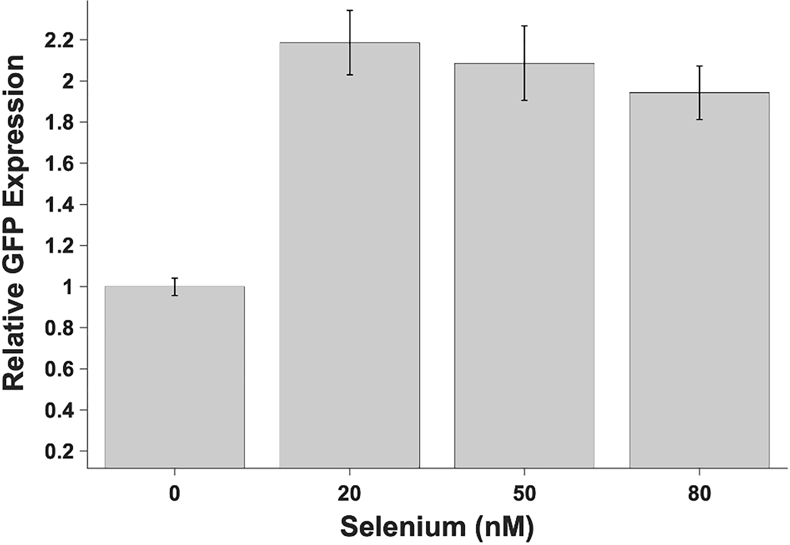
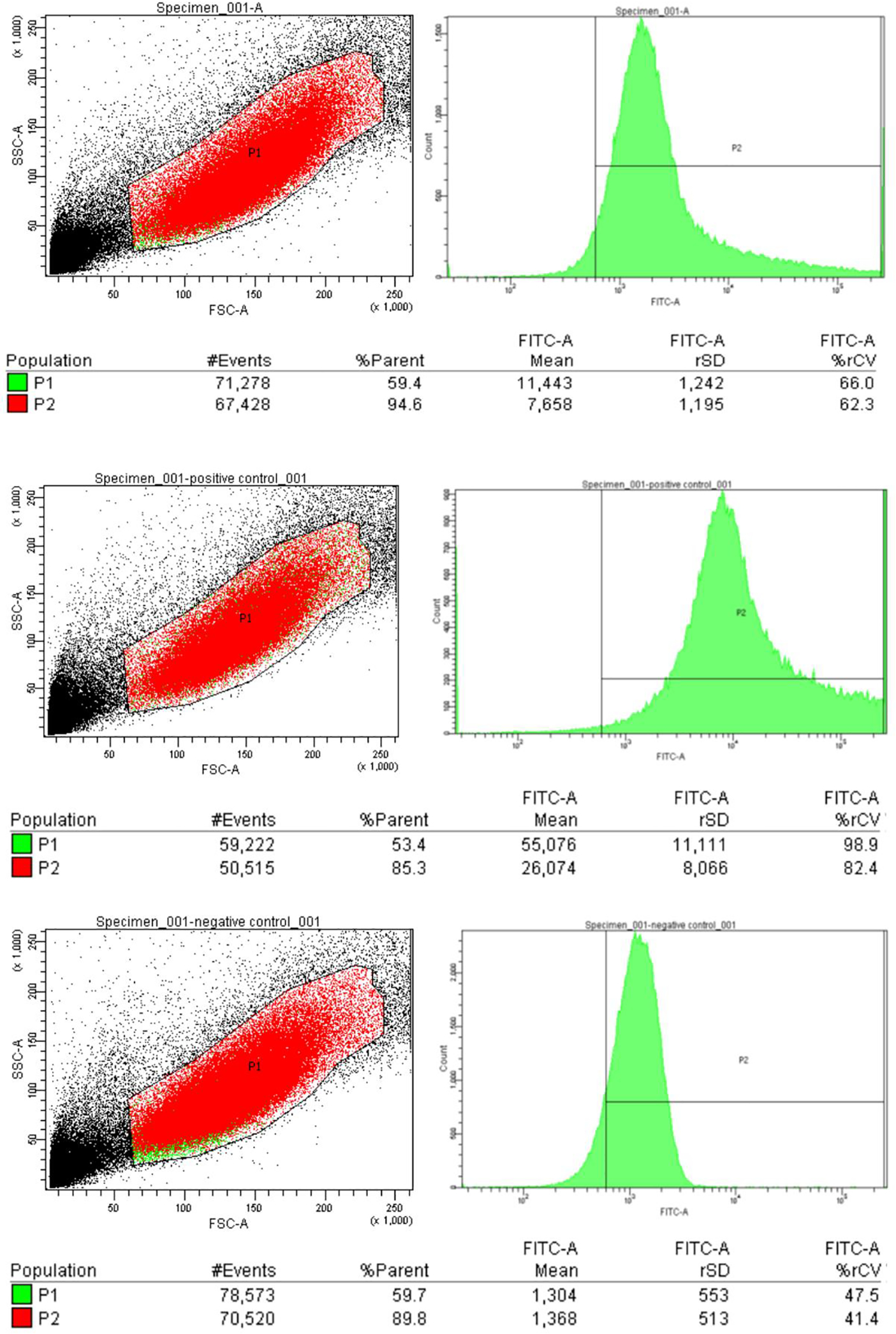
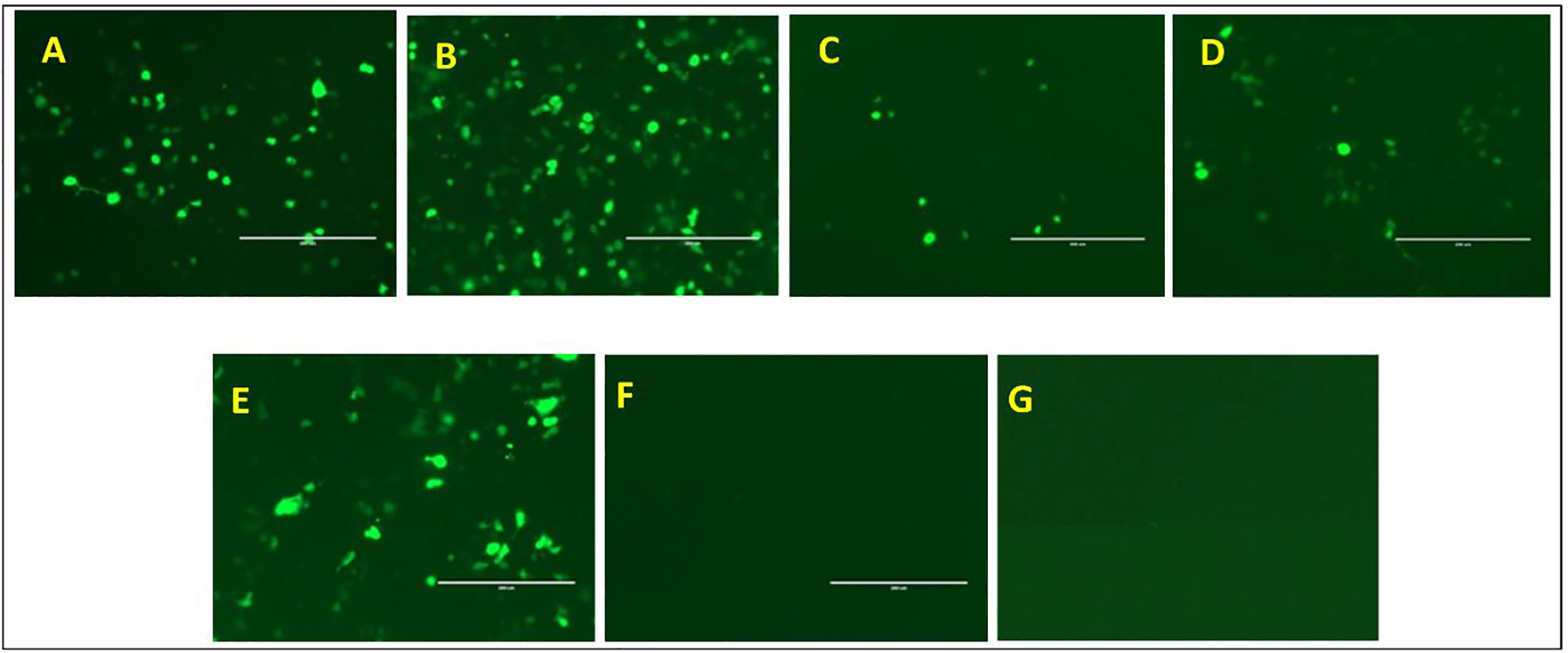
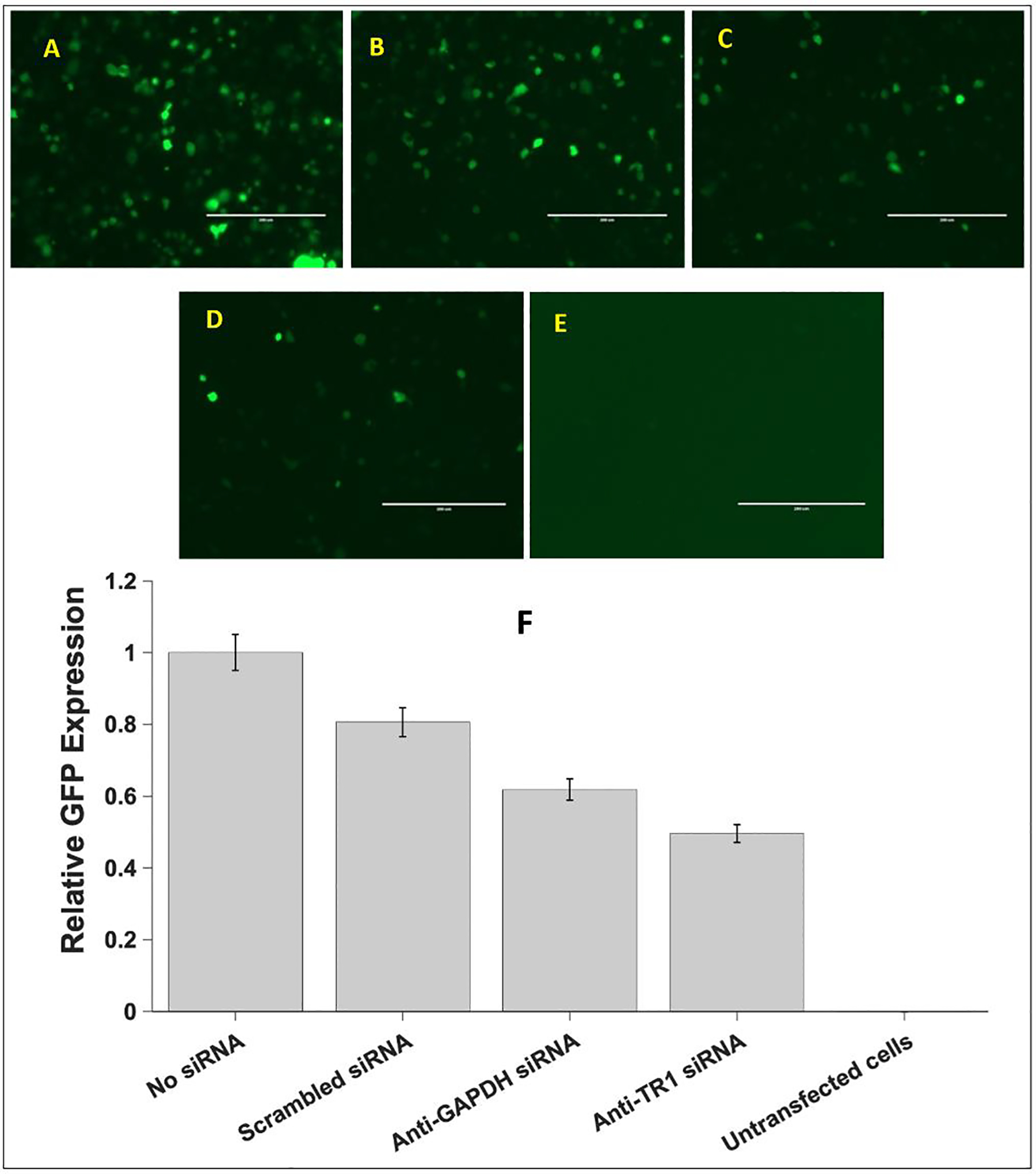
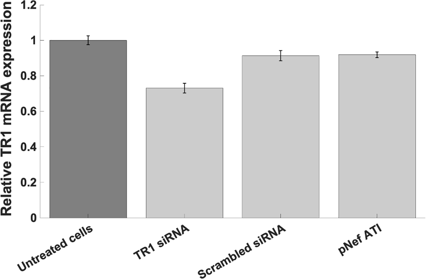
The HIV-1 nef gene terminates in a 3'-UGA stop codon, which is highly conserved in the main group of HIV-1 subtypes, along with a downstream potential coding region that could extend the nef protein by 33 amino acids, if readthrough of the stop codon occurs. Antisense tethering interactions (ATIs) between a viral mRNA and a host selenoprotein mRNA are a potential viral strategy for the capture of a host selenocysteine insertion sequence (SECIS) element (Taylor et al, 2016) [1]. This mRNA hijacking mechanism could enable the expression of virally encoded selenoprotein modules, via translation of in-frame UGA stop codons as selenocysteine (SeC). Here we show that readthrough of the 3'-terminal UGA codon of nef occurs during translation of HIV-1 nef expression constructs in transfected cells. This was accomplished via fluorescence microscopy image analysis and flow cytometry of HEK 293 cells, transfected with engineered GFP reporter gene plasmid constructs, in which GFP can only be expressed by translational recoding of the UGA codon. SiRNA knockdown of thioredoxin reductase 1 (TR1) mRNA resulted in a 67% decrease in GFP expression, presumably due to reduced availability of the components involved in selenocysteine incorporation for the stop codon readthrough, thus supporting the proposed ATI. Addition of 20 nM sodium selenite to the media significantly enhanced stop codon readthrough in the pNefATI1 plasmid construct, by >100%, supporting the hypothesis that selenium is involved in the UGA readthrough mechanism.


Conflict of interest statement

Conflicts of Interest All authors declare that they have no conflicts of interest.

Figures

Fig. 1.
Fig. 1.. Proposed mechanism of Sec incorporation into viral proteins via hijacking of a SECIS element from a tethered host selenoprotein mRNA.
(Reproduced from Fig. 3 of Taylor et al, 2016 [1]). Both panels show schematic ribosomes with two bound tRNAs, one carrying the Sec, the other a growing peptide chain shown as colored circles. The upper panel shows the established role of the SECIS element in the 3’-UTR region in the mechanism of insertion of Sec during mammalian selenoprotein biosynthesis [5,10]. The panel below shows how the HIV-1 mRNA could hijack the host SECIS element via antisense tethering interactions (ATI) to decode UGA to synthesize viral selenoproteins such as the HIV-1 encoded GPx [7]. ATI-1 is a predicted interaction spanning the highly conserved 3’ UGA codon of the nef gene. ATI-2 is a second shorter antisense region consisting of 13 consecutive bases near the end of viral mRNA.
Fig. 2.
Fig. 2.. Schematics for the plasmid inserts used to assess nef 3’-UGA readthrough.
Regions with antisense complementarity to TR1 mRNA are shown crosshatched as ATI1 and ATI2. Naturally occurring (TGA, TAA) or engineered (TAG) in-frame stop codons and a CAA mutant of the wild-type HIV TAA are indicated.
Fig. 3.
Fig. 3.. Readthrough of the HIV-1 nef 3’-UGA codon .
The photomicrograph panels A-E show GFP fluorescence in HEK 293T cells, transfected with three different ATI plasmid vector constructs and controls. (A) pNefATI1; (B) pNefATI2; (C) pNefATIstop; (D) Parent GFP construct; (E) Untransfected cells; (F) Bar graph showing the GFP intensity of each transfection condition calculated using NIH ImageJ Software.
Fig 4.
Fig 4.. Added selenium enhances stop codon readthrough from the ATI-1 plasmid construct.
Results show that supplementation of the basal media with additional selenium as sodium selenite has a significant effect on the stop codon readthrough, resulting in higher levels of GFP expression. However, the maximal readthrough was observed at the lowest level of selenium (20nM) and the readthrough did not change significantly at higher selenium concentrations. Even at the 20 nM concentration, addition of sodium selenite essentially doubles UGA stop codon readthrough relative to unmodified cell culture medium, as measured by GFP production.
Fig. 5.
Fig. 5.. Flow cytometry analysis of HIV-1 nef stop codon readthrough.
A (Top): HEK 293T Cells transfected with pNefATI1 vector. FITC-A represents green fluorescence (GFP). The P1 population had a mean FITC-A of 11,443 ± 1,242. B (Middle): Cells transfected with EGFP-N3 plasmid. The P1 population had a mean FITC-A of 55,076 ± 11,111. C (Bottom): Untransfected cells (background). The mean FITCA of P1 was 1,304 ± 553. Stop codon readthrough efficiency was thus 18.9% (see text).
Fig. 6.
Fig. 6.. Selection of transfection reagent and optimization of the transfection reagent volume using HEK 293T cells transfected with pNefATI1 EGFP-N3 vector.
EVOS GFP fluorescence images A, B, C, D, E and F are cells transfected with 0.3 ul amine, 0.6 ul amine, 0.15 ul amine, 0.5 ul NeoFX, 1.2 ul NeoFX and 0.15 ul NeoFx respectively. Image G represents untransfected cells.
Fig 7.
Fig 7.. Effect of siRNAs on GFP production from the pNefATI1 construct.
Panels A-E show EVOS microscopy images of each transfection condition: A. pNefATI1 with no siRNA. B. With added negative control scrambled siRNA. C. With positive control anti-GAPDH siRNA. D. With anti-TR1 siRNA. E. Untransfected cells. F. Bar graph showing average GFP expression from 10 images of each treatment calculated using NIH ImageJ software.
Fig 8.
Fig 8.. TR1 mRNA expression assessed by qPCR.
Compared to untreated cells, an approximately 27% knockdown of TR1 mRNA relative to untreated cells (P < 0.0001) was observed in the sample treated with anti-TR1 siRNA. This confirms the knockdown of TR1 mRNA that may contribute to the decrease in nef 3’-UGA readthrough in the presence of anti-TR1 siRNA (Fig. 7).

Similar articles

Cited by

References

    1. Taylor EW; Ruzicka JA; Premadasa L; Zhao L Cellular selenoprotein mRNA tethering via antisense interactions with Ebola and HIV-1 mRNAs may impact host selenium biochemistry. Current Topics in Medicinal Chemistry 2016, 16, 1530–1535. - PMC - PubMed
    1. Zhong L; Arner ES; Holmgren A Structure and mechanism of mammalian thioredoxin reductase: the active site is a redox-active selenolthiol/selenenylsulfide formed from the conserved cysteine-selenocysteine sequence. Proc Natl Acad Sci U S A 2000, 97, 5854–5859, doi:10.1073/pnas.100114897. - DOI - PMC - PubMed
    1. Gonzalez-Flores JN; Shetty SP; Dubey A; Copeland PR The molecular biology of selenocysteine. Biomolecular concepts 2013, 4, 349–365, doi:10.1515/bmc-2013-0007. - DOI - PMC - PubMed
    1. Taylor EW; Cox AG; Zhao L; Ruzicka JA; Bhat AA; Zhang W; Nadimpalli RG; Dean RG Nutrition, HIV, and drug abuse: the molecular basis of a unique role for selenium. J Acquir Immune Defic Syndr 2000, 25 Suppl 1, S53–61. - PubMed
    1. Donovan J; Copeland PR Evolutionary history of selenocysteine incorporation from the perspective of SECIS binding proteins. BMC evolutionary biology 2009, 9, 229, doi:10.1186/1471-2148-9-229. - DOI - PMC - PubMed

LinkOut - more resources