. 2022 Feb;13(2):4010-4027.
doi: 10.1080/21655979.2022.2031411.

DNA methyltransferase 3 beta regulates promoter methylation of microRNA-149 to augment esophageal squamous cell carcinoma development through the ring finger protein 2/Wnt/β-catenin axis

Affiliations

DNA methyltransferase 3 beta regulates promoter methylation of microRNA-149 to augment esophageal squamous cell carcinoma development through the ring finger protein 2/Wnt/β-catenin axis

Junfeng Yang et al. Bioengineered. 2022 Feb.

Abstract

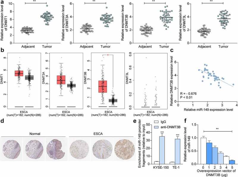
Esophageal squamous cell carcinoma (ESCC) is an aggressive form of human squamous cell carcinomas with extremely aggressive pathological features. This study explores the functions of microRNA-149 (miR-149) and its interacted molecules in ESCC. The ESCC-related miRNA and messenger RNA (mRNA) datasets were applied to identify aberrantly expressed genes in ESCC. Forty-two patients with ESCC were included and their tissue samples were collected. miR-149 was poorly expressed whereas DNA methyltransferase 3 beta (DNMT3B) and ring finger protein 2 (RNF2) were abundantly expressed in ESCC tumor samples. Overexpression of miR-149 suppressed growth and invasiveness of ESCC cells in vitro and in vivo. DNMT3B bound to the promoter region of miR-149 to trigger its promoter methylation and downregulation. RNF2 mRNA was a target of miR-149. RNF2 overexpression blocked the inhibitory effect of miR-149 on ESCC cell growth. RNF2 activated the Wnt/β-catenin pathway to promote ESCC development. In conclusion, this study found that DNMT3B downregulates miR-149 level through methylation modification of the miR-149 promoter, while miR-149 suppresses RNF2 expression and inactivates the Wnt/β-catenin pathway to suppress growth of ESCC cells.

Keywords: DNMT3B; Esophageal squamous cell carcinoma; RNF2; methylation; miR-149; wnt/β-catenin.


Conflict of interest statement

No potential conflict of interest was reported by the author(s).

Figures

None
Graphical abstract
Figure 1.
Figure 1.
miR-149 is expressed at low levels in ESCC tissues and cells. A, DE miRNAs between ESCC tumor and normal tissues screened using a GEO GSE67268 dataset; B, miR-149 expression in ESCC and the paired adjacent tissue samples from 42 patients detected by RT-qPCR; C-E, association between miR-149 and the clinical staging (c), lymph node metastasis (d) and tumor differentiation (e); F, miR-149 expression in ESCC cell lines (TE-1, EC9706 and KYSE-150) and in HET-1A cells determined by RT-qPCR. In panels B-E, each plot indicates a single sample. **p < 0.01.
Figure 2.
Figure 2.
miR-149 suppresses malignance of ESCC cells in vitro. a, expression of miR-149 in KYSE-150 and TE-1 cells after miR-149 mimic or mimic control transfection detected by RT-qPCR; b, colony formation ability of cells; c, Caspase-3 activity in cells detected by a Caspase-3 kit; d, apoptosis rate in cells determined by flow cytometry; e, mRNA levels of the EMT-related factors (E-cadherin, ZO-1, N-cadherin and Vimentin) in ESCC cells measured by RT-qPCR; f-g, migration (f) and invasiveness (g) of cells evaluated by Transwell assays. **p < 0.01.
Figure 3.
Figure 3.
miR-149 suppresses growth of ESCC cells in vivo. a, weekly change in the volume of xenograft tumors; b, tumor weight on the 36th day; c-d, expression of PCNA (c) and KI67 (d) in tumor examined by IHC; E, cell apoptosis rate in tumor tissues determined by TUNEL assay. N = 6 in each group. **p < 0.01.
Figure 4.
Figure 4.
The promoter region of miR-149 is highly methylated in ESCC. a, location of miR-149 and GPC1 in genome measured using the Ensembl system; b, the CpG Island on the miR-139 promoter predicted using the UCSC browser; c, promoter methylation level of miR-149 in tumor and healthy tissues examined by MSP-qPCR; d, correlation between the promoter methylation level of miR-149 and its expression in tumor tissues; e, promoter methylation level of miR-149 in ESCC cell lines (TE-1, EC9706 and KYSE-150) and in HET-1A cells determined by MSP-qPCR; f, miR-149 expression in TE-1, EC9706 and KYSE-150 cells after 5-aZa-CDR treatment determined by RT-qPCR. In panels C and D, each spot indicates a single sample. **p < 0.01.
Figure 5.
Figure 5.
DNMT3B regulates methylation of miR-149 promoter. a, DNMT1, DNMT3A, DNMT3B and DNMT3L mRNA levels in ESCC and normal tissues from 42 collected patients detected by RT-qPCR; b, DNMT1, DNMT3A, DNMT3B and DNMT3L expression predicted in TCGA-ESCA; c, an inverse correlation of miR-149 and DNMT3B in ESCC tissues; d, strong positive staining of DNMT3B in ESCC; E-F, binding between DNMT3B and miR-149 promoter verified through ChIP-qPCR (e) and luciferase (f) assays. In panels a and c, each spot indicates a sample. **p < 0.01.
Figure 6.
Figure 6.
miR-149 directly targets RNF2 mRNA. a-b, DE genes between ESCC and normal tissues analyzed using the GEO GSE29001 and GSE20347 datasets; c, a Venn diagram for target mRNAs of miR-149 predicted via bioinformatics systems and DE genes screened from two datasets; d, RNF2 mRNA in ESCC and normal tissues detected by RT-qPCR; E-F, correlation of RNF2 expression with miR-149 (e) and DNMT3B (f) in ESCC tumor tissues; g, predicted RNF2 expression in TCGA-ESCA; H, staining intensity of RNF2 in ESCC and normal tissues examined by the IHC assay; i, mRNA expression of RNF2 in ESCC cells and in HET-1A cells determined by RT-qPCR; j, binding between miR-149 and RNF2 verified by luciferase assay; k, RNF2 mRNA expression in TE-1 and KYSE-150 cells after miR-149 mimic transfection determined by RT-qPCR. In panel D-F, each spot indicates a sample. **p < 0.01.
Figure 7.
Figure 7.
Overexpression of RNF2 promotes ESCC cell growth and metastasis. a, RNF2 mRNA in KYSE-150 and TE-1 cells after oe-RNF-2 administration examined by RT-qPCR; b, colony formation ability of cells; c, Caspase-3 activity in cells determined using a Caspase-3 kit; d, apoptosis of cells detected by flow cytometry; E-F, migration (e), and invasiveness (f) of cells measured by Transwell assays. **p < 0.01.
Figure 8.
Figure 8.
RNF2 activates the Wnt/β-catenin pathway. a, the RNF2-related signaling pathways predicted using GSEA; b, protein levels of Wnt1 and β-catenin in ESCC cells detected by Western blot analysis; c, sub-cellular localization of β-catenin in cells examined by immunofluorescence staining. **p < 0.01.
Figure 9.
Figure 9.
A graphic abstract. DNMT3B induces CpG Island methylation at the promoter of miR-149 on the GPC1 transcript, which results in miR-149 downregulation and restoration of RNF2 mRNA, leading to Wnt/β-catenin pathway activation and augmented development of ESCC.

Similar articles

Cited by

References

    1. Rustgi AK, El-Serag HB, Ingelfinger JR.. Esophageal carcinoma. N Engl J Med. 2014;371(26):2499–2509. - PubMed
    1. Tirumani H, Rosenthal MH, Tirumani SH, et al. Esophageal carcinoma: current concepts in the role of imaging in staging and management. Can Assoc Radiol J. 2015;66(2):130–139. - PubMed
    1. Wang J, Zhu W, Tao G, et al. Circular RNA circ-LRP6 facilitates Myc-driven tumorigenesis in esophageal squamous cell cancer. Bioengineered. 2020;11(1):932–938. - PMC - PubMed
    1. Domper Arnal MJ, Ferrandez Arenas A, Lanas Arbeloa A. Esophageal cancer: risk factors, screening and endoscopic treatment in Western and Eastern countries. World J Gastroenterol. 2015;21(26):7933–7943. - PMC - PubMed
    1. Adams BD, Kasinski AL, Slack FJ. Aberrant regulation and function of microRNAs in cancer. Curr Biol. 2014;24(16):R762–76. - PMC - PubMed

Publication types

MeSH terms