. 2023 Mar 1;277(3):e634-e647.
doi: 10.1097/SLA.0000000000005252. Epub 2021 Oct 12.

Pseudomonas Aeruginosa Theft Biofilm Require Host Lipids of Cutaneous Wound

Affiliations

Pseudomonas Aeruginosa Theft Biofilm Require Host Lipids of Cutaneous Wound

Mithun Sinha et al. Ann Surg. .

Abstract

Objective: This work addressing complexities in wound infection, seeks to test the reliance of bacterial pathogen Pseudomonas aeruginosa (PA) on host skin lipids to form biofilm with pathological consequences.

Background: PA biofilm causes wound chronicity. Both CDC as well as NIH recognizes biofilm infection as a threat leading to wound chronicity. Chronic wounds on lower extremities often lead to surgical limb amputation.

Methods: An established preclinical porcine chronic wound biofilm model, infected with PA or Pseudomonas aeruginosa ceramidase mutant (PA ∆Cer ), was used.

Results: We observed that bacteria drew resource from host lipids to induce PA ceramidase expression by three orders of magnitude. PA utilized product of host ceramide catabolism to augment transcription of PA ceramidase. Biofilm formation was more robust in PA compared to PA ∆Cer . Downstream products of such metabolism such as sphingosine and sphingosine-1-phosphate were both directly implicated in the induction of ceramidase and inhibition of peroxisome proliferator-activated receptor (PPAR)δ, respectively. PA biofilm, in a ceram-idastin-sensitive manner, also silenced PPARδ via induction of miR-106b. Low PPARδ limited ABCA12 expression resulting in disruption of skin lipid homeostasis. Barrier function of the wound-site was thus compromised.

Conclusions: This work demonstrates that microbial pathogens must co-opt host skin lipids to unleash biofilm pathogenicity. Anti-biofilm strategies must not necessarily always target the microbe and targeting host lipids at risk of infection could be productive. This work may be viewed as a first step, laying fundamental mechanistic groundwork, toward a paradigm change in biofilm management.


Conflict of interest statement

The authors report no conflict of interest.

Figures
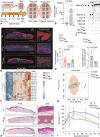
Figure 1
Figure 1
Inducible Bacterial Ceramidases Deplete Host Cutaneous Ceramide. (A) Schematic presentation of the timeline of porcine model of chronic wound biofilm infection. (B) Induction of bacterial ceramidase (bcdase) in biofilm infected porcine wounds. Real-time qPCR analysis of bcdase expression spontaneously infected (SI) and wild type P. aeruginosa (PAwt) infected porcine wound epidermis (d7 post-infection) collected by laser capture micro-dissection (LCM). Data presented as mean±SEM, (n=5). (C) PA∆cer biofilm was deficient in secreted bcdase activity. Data presented as mean±SEM, (n=6). See Fig. S1A, http:// links.lww.com/SLA/D496 for gene bcdase expression data. [NBD-CER - C12-NBD-Ceramide, NBD-DA - NBD-dodecanoic acid]. (D and E) Depletion of ceramide in porcine wounds infected with PA wt biofilm compared to SI or wounds infected with ceramidase mutant of P. aeruginosa (PAΔcer). Porcine d56 wound tissues were immuno-stained with anti-ceramide (red) antibody and DAPI (blue). Data presented as mean±SEM, (n=6). Scale bar=500 µm and zoomed inset=50 µm. (F) Depletion of ceramides in porcine skin lipid exposed to PA wt ,or PAΔcer as measured by LC/MS/MS targeted-lipidomic approach. Same skin lipid not Exposed to any bacteria was used as sham control. Hierarchical cluster analysis revealed down-regulation of 19 species of ceramide in the skin lipid (N=6). Individual ceramides that were specifically affected are illustrated in Supplementary Fig. S4, http:// links.lww.com/sla/d496. (G) Principal component analyses of the abundance of cutaneous ceramides in response to PAWT or PAΔCER exposure. Sham and PAΔCER clusters were not statistically different. This combined group was statistically distinct from cutaneous ceramide levels in response to PAWT. (H) Complete re-epithelization following SI, PAWT and PAΔCER biofilm infection. Hematoxylin-eosin staining shown. Scale bar=500 μm and zoomed inset=50 μm. Macroscopic digital planimetry shown in Supplementary Fig. S5M, http://links.lww.com/sla/d496. (I) functional restoration of Barrier function of cutaneous wounds, measured by tewl, was compromised following PAWT biofilm infection. Data presented as mean±SEM (N=6). / P<0.05 as compared to PAΔCER; and *P<0.05 as compared to SI.
Figure 2
Figure 2
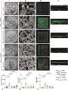
Host Lipids Facilitate PAwt Biofilm Aggregate Formation. (A-E) (i) Cutaneous lipids induced biofilm aggregate formation in an in vitro polycarbonate membrane biofilm system after 24 h of inoculation. A, PAwt +vehicle (PBS); B, PAwt + skin lipids; C, PAwt + depleted lipids; D, PA∆cer +vehicle (PBS) E, PA∆cer + skin lipids. (ii) Increased abundance of EPS in PAwt biofilm in response to host lipids as recorded in SEM images. Thick EPS is marked by yellow circles. EPS fibers are marked by yellow arrows. Scale bar = 100 nm, 30,000x magnification. Larger SEM fields are presented as supplementary Figure 6, http://links.lww.com/SLA/D496. (iii) Host lipids induced biofilm aggregate formation in PAwt as measured by wheat germ agglutinin (WGA) staining. Scale bar = 10 µm. (iv) Respective 3D reconstructed images of (iii). (F) Quantification of biofilm aggregates using WGA staining as shown in (iii). Data presented as mean ± SEM, n= 10. (G-H) Host lipids induce biofilm gene expression in pathogen PAwt. Data presented as mean ± SEM, (n = 5).
Figure 3
Figure 3
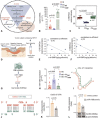
Biogenesis of host long chain ceramides is compromised under conditions of PAwt Biofilm infection. (A) Long chain ceramide biogenesis pathways outlined. (B) Downregulation of CerS3 in wound-site skin tissue infected with PAwt compared to SI or PA∆cer measured by qPCR. Data presented as mean ± SEM (n = 5-7). (C) PAwt lowered the levels of dihydroceramide (C18DHCer) in skin lipids. Measured by LC/MS/MS targeted lipidomic approach. Data are presented as mean ± SEM (n = 6). (D) Wound fluid collection from chronic wound patients who underwent negative pressure wound therapy (NPWT) as part of standard of care. (E and F) Lower levels of dihydroceramide in cyclic diGMP-rich human wound fluid. cyclic di-GMP is a marker of PA biofilm infection (n = 5). (G) Biofilm induced miR-106b targets host CerS3 (H) Elevated miR-106b in human keratinocytes (HK) infected with PAwt biofilm as measured by qPCR. Data presented as mean ± SEM (n = 7–10). (I and J) miR-106b is predicted to target the 3’–UTR of CerS3 position 331–355 according to RNAHybrid algorithm. (K) miR-106b silenced pmiR-Target-Cers–3’–UTR in human keratinocytes (HK). FL indicates Firefly luciferase;RL, Renilla luciferase. Data are mean ± SEM (n = 4). (L) miR-106b silenced CerS3 protein expression in human keratinocytes (HK). Data are presented as mean ± SEM (n = 6).
Figure 4
Figure 4
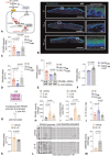
Wound-Site Skin PPARδ Expression is Downregulated in Response to PA^t Biofilm Infection. (A) PAwt biofilm compromises host cutaneous PPARδ expression. (B and C) Loss of cutaneous host PPARB in porcine wounds infected with PAwt compared to SI or PA∆cer wounds. Porcine wound sections were immunostained with anti-PPARδ (green) and DAPI (blue). Porcine wound section images (scale bar = 500 µm) and corresponding zoomed inset (scale bar = 50 µm) showed PPARδ protein expression. Data presented as mean ± SEM (n = 5–6). (D) Lower expression of PPARδ in PAwt infected wound tissue. Whole tissue homogenate was used for analysis. Data presented as mean ± SEM, (n = 3–8). (E) Lower expression of PPARδ protein in human keratinocytes (HK) infected with PAwt biofilm. Data presented as mean ± SEM (n = 5). (F) Ceramidastin attenuated the negative effects of PAwt biofilm on PPARB expression in human keratinocytes. Ceramidastin, 10 µg/mL. Data presented as mean ± SEM (n = 6). (G) PPARB promoter assay approach. (H) Increased PPARδ transactivity in human keratinocytes (HK) treated with long chain ceramides (C18 & C24, 5 µmol/mL). Nuclear extract of the transfected cells treated for 48 h were used to measure PPARB trans-activity. Data presented as mean ± SEM (n = 5). (I) Activation of PPARδ promoter by long chain ceramides as measured by reporter assay. Human keratinocytes (HK) were transfected with PPARB promoter along with long chain ceramides (C:18, C:24, 5 µmol/mL). Data presented as mean ± SEM (n = 3). (J) Elevated levels of sphingosine-1-phosphate (S-1-P) in PAwt infected wound tissues. Measured by LC/MS/MS targeted lipidomic approach. Cutaneous wound tissue infected with PAwt showed higher levels of S-1-P as compared to SI or PA∆cer infected wounds. Data presented as mean ± SEM (n = 6). (K) Down-regulation of PPARδ gene expression in human keratinocytes (HK) receiving S-1-P (5 µmol/mL). Data presented as mean ± SEM (n = 6-7). (L) Region of PPARδ promoter analyzed through bisulfite genomic sequencing of DNA. Methylation profile of the PPARB promoter in human keratinocytes (HK) treated with S-1-P (5 µmol, 48 h). Methylated CpG in black (filled) and unmethylated CpG in white (open). Number of clones = 10. (M) Increased DNA methyl transferase 3B (DNMT3B) activity in human keratinocytes (HK) treated S-1-P (5 µmol/L, 48h). Data presented as mean ± SEM (n = 4-5).
Figure 5
Figure 5
miR-106b Targets PPARB in PAwt Biofilm Infected Keratinocytes. (A) RNA HybridTM-based prediction shows that PPARB 3’–UTR isa target of miR-106b. (B) PPARB downregulation in miR-106b mimic transfected human keratinocytes (HK) as measured by qPCR. Data presented as mean ± SEM (n = 3-4). (C) Downregulated PPARB protein in response to miR-106b mimic as measured by Western blot in HK. Data presented as mean ± SEM (n = 6). (D) miR-106B targets PPARB 3’UTR as shown by reporter assay. HK were transfected with HMIT013627 -MT06 - PPARΔ -3’-UTR (NM_006238) along with miR-106B mimic or control mimic. Data presented as mean ± SEM (N = 5).
Figure 6
Figure 6
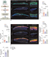
ABCA12 Expression Is Compromised Following PAwt Biofilm Infection. (A) Schematic depiction of the hypothetical pathway. (B and C) Loss of ABCA12 in wound-site epithelium which was infected with PAwt compared to SI or PA∆cer groups. Scale bar = 500 µm with corresponding zoomed images of 50 µm. anti-ABCA12 (green) and DAPI (blue). Data presented as mean ± SEM (n = 5). (D) Downregulation of ABCA12 in porcine wounds infected with PAwt compared to SI or PA∆cer wounds measured by qPCR. Whole tissue homogenate was used for analysis. Data presented as mean ± SEM (n = 5–8). (E) Disruption of neutral and polar lipid distribution at the affected wound-site skin. Scale bar = 500 µm with corresponding zoomed images of 50 µm. Nile Red staining with DAPI (blue) showing expression of polar lipids (red) and neutral lipids (green). Data presented as mean ± SEM (n = 5). (F) Ceramidastin (10 µg/mL) attenuated biofilm-induced loss of ABCA12 expression in human keratinocytes (HK) as measured by qPCR. Data presented as mean ± SEM (n = 5-6).
Figure 7
Figure 7
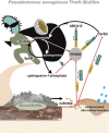
Summary figure. In cutaneous wounds, Pseudomonas aeruginosa forms pathogenic theft biofilm. Such biofilm severity relies on the theft of host lipids (ceramides) causing potent induction of bacterial ceramidase (bcdase). Skin ceramide biosynthesis is impaired by post-trancriptional gene silencing of CerS3 by biofilm-inducible miR106b. The limited ceramide pool, thus available as inducer of PPARδ, is threatened by elevated biofilm bcdase. Thus expression of PPARδ, a major regulator of skin lipid homeostasis, is blunted. PPARδ faces a second line of attack in the form of epi-genetic silencing by both biofilm-induc-ible miR–106b as well as S-1-P. Such loss of PPARδ compromised downstream genes including cutaneous lipid transporter ABCA12. Taken together, biofilm disrupts skin lipid homeostasis in a way that the site of wound repair cannot restore barrier function, a marker of functional wound healing.

References

    1. Barki KG, Das A, Dixith S, et al. Electric field based dressing disrupts mixed-species bacterial biofilm infection and restores functional wound healing. Ann Surg. 2019;269:756–766. - PMC - PubMed
    1. Roy S, Elgharably H, Sinha M, et al. Mixed-species biofilm compromises wound healing by disrupting epidermal barrier function. J Pathol. 2014;233:331–343. - PMC - PubMed
    1. Roy S, Santra S, Das A, et al. Staphylococcus aureus biofilm infection compromises wound healing by causing deficiencies in granulation tissue collagen. Ann Surg. 2020;271:1174–1185. - PMC - PubMed
    1. Wolcott R, Dowd S. The role of biofilms: are we hitting the right target? Plast Reconstr Surg. 2011;127(suppl 1):28S–35S. - PubMed
    1. Fazli M, Bjarnsholt T, Kirketerp-Moller K, et al. Nonrandom distribution of Pseudomonas aeruginosa and Staphylococcus aureus in chronic wounds. J Clin Microbiol. 2009;47:4084–4089. - PMC - PubMed

Publication types