

New imaging tools for mouse models of osteoarthritis
- PMID: 35129777
- PMCID: PMC9135906
- DOI: 10.1007/s11357-022-00525-3
New imaging tools for mouse models of osteoarthritis
Abstract
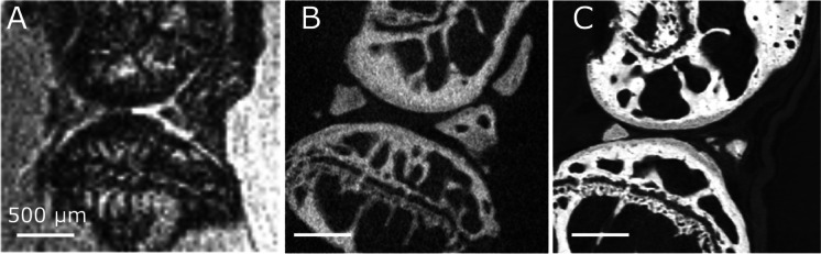
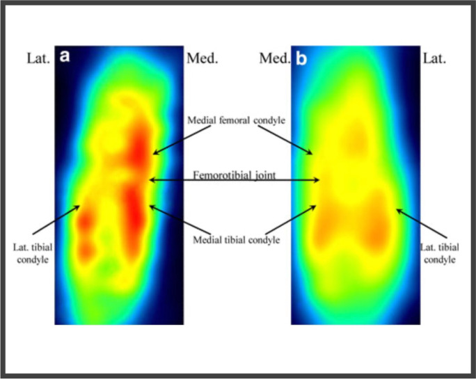
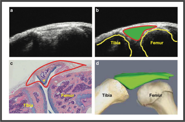
Osteoarthritis (OA) is a chronic degenerative disease characterized by a disruption of articular joint cartilage homeostasis. Mice are the most commonly used models to study OA. Despite recent reviews, there is still a lack of knowledge about the new development in imaging techniques. Two types of modalities are complementary: those that assess structural changes in joint tissues and those that assess metabolism and disease activity. Micro MRI is the most important imaging tool for OA research. Automated methodologies for assessing periarticular bone morphology with micro-CT have been developed allowing quantitative assessment of tibial surface that may be representative of the whole OA joint changes. Phase-contrast X-ray imaging provides in a single examination a high image precision with good differentiation between all anatomical elements of the knee joint (soft tissue and bone). Positron emission tomography, photoacoustic imaging, and fluorescence reflectance imaging provide molecular and functional data. To conclude, innovative imaging technologies could be an alternative to conventional histology with greater resolution and more efficiency in both morphological analysis and metabolism follow-up. There is a logic of permanent adjustment between innovations, 3R rule, and scientific perspectives. New imaging associated with artificial intelligence may add to human clinical practice allowing not only diagnosis but also prediction of disease progression to personalized medicine.
Keywords: Imaging; Innovation; Mouse models; Osteoarthritis; Technologies.
© 2022. The Author(s), under exclusive licence to American Aging Association.
Conflict of interest statement
The authors declare no competing interests.
Figures

References
-
- GBD Disease and injury incidence and prevalence collaborators (2017) global, regional, and national incidence, prevalence, and years lived with disability for 328 diseases and injuries for 195 countries, 1990–2016: a systematic analysis for the global burden of disease study 2016. Lancet. 2016;390:1211–1259. doi: 10.1016/S0140-6736(17)32154-2. - DOI - PMC - PubMed
-
- Buckwalter JA, Mankin HJ. Articular cartilage: tissue design and chondrocyte-matrix interactions. Instr Course Lect. 1998;47:477–486. - PubMed
Publication types
MeSH terms
LinkOut - more resources
Full Text Sources
