

Bovine rhinitis B virus is highly prevalent in acute bovine respiratory disease and causes upper respiratory tract infection in calves
- PMID: 35130139
- PMCID: PMC8941992
- DOI: 10.1099/jgv.0.001714
Bovine rhinitis B virus is highly prevalent in acute bovine respiratory disease and causes upper respiratory tract infection in calves
Abstract
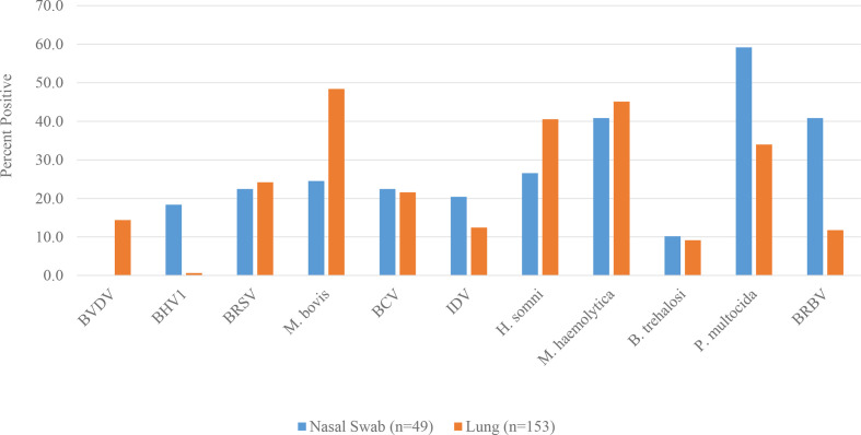
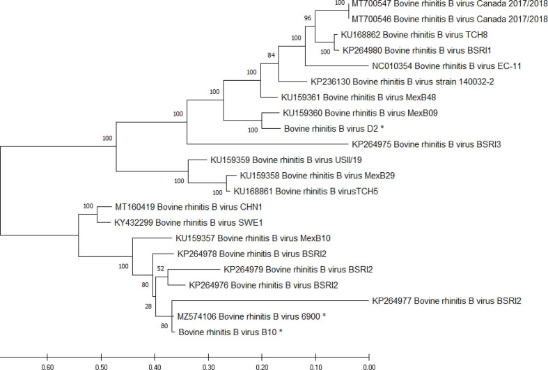
Bovine respiratory disease (BRD) is the most significant cause of cattle morbidity and mortality worldwide. This multifactorial disease has a complex aetiology. Dogma posits a primary viral infection followed by secondary bacterial pneumonia. Bovine rhinitis B virus (BRBV) is an established aetiological agent of BRD, but little is known regarding its pathogenesis. Here, a BRD PCR panel identified 18/153 (11.8 %) lung samples and 20/49 (40.8 %) nasal swabs collected from cattle with respiratory signs as positive for BRBV, which was the most prevalent virus in nasal swabs. Primary bovine tracheal epithelial cells were used to isolate BRBV that was phylogenetically related to contemporary sequences from the USA and Mexico and genetically divergent from the previous sole BRBV isolate. To investigate virus pathogenesis, 1-week-old colostrum-deprived dairy calves were inoculated intranasally with 7.0 log10 TCID50 BRBV. Virus was isolated from nasal swabs, nasal turbinates, trachea and the brain of the challenged animals. Neutralizing antibodies were detected beginning 7 days post-inoculation and peaked at day 14. In situ hybridization (ISH) localized BRBV infection in the upper respiratory ciliated epithelial and goblet cells, occasionally associated with small defects of the superficial cilia lining. Sporadically, pinpoint ISH signals were also detected in cells resembling glial cells in the cerebrum in one calf. Together, these results demonstrate the BRBV infection is highly prevalent in acute BRD samples and while the pathogenicity of BRBV is minimal with infection largely limited to the upper respiratory tract, further research is needed to elucidate a possible initiatory role in BRD.
Keywords: bovine respiratory disease; bovine rhinitis virus; picornavirus.
Conflict of interest statement
The authors declare that there are no conflicts of interest.
Figures


References
-
- Mitra N, Cernicchiaro N, Torres S, Li F, Hause BM. Metagenomic characterization of the virome associated with bovine respiratory disease in feedlot cattle identified novel viruses and suggests an etiologic role for influenza D virus. J Gen Virol. 2016;97:1771–1784. doi: 10.1099/jgv.0.000492. - DOI - PMC - PubMed
-
- Bögel K, Böhm H. Ein rhinovirus des rindes. Zentralbl Bakteriol Orig. 1962;193:2–14.
Publication types
MeSH terms
LinkOut - more resources
Full Text Sources