Review
. 2022 Jan 25;54(1):25-36.
doi: 10.3724/abbs.2021003.

Bacterial MerR family transcription regulators: activationby distortion

Affiliations
Review

Bacterial MerR family transcription regulators: activationby distortion

Chengli Fang et al. Acta Biochim Biophys Sin (Shanghai). .

Abstract

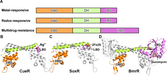
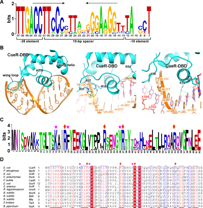
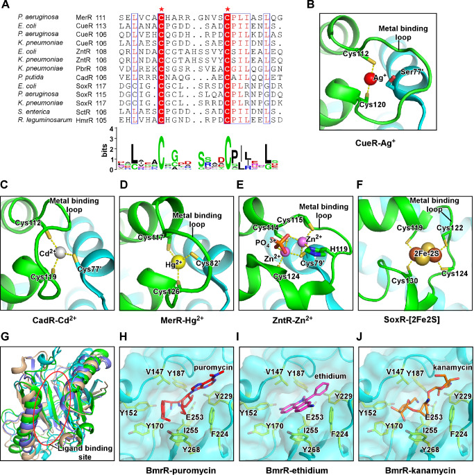
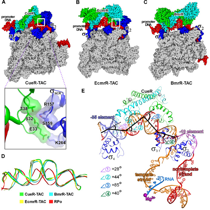
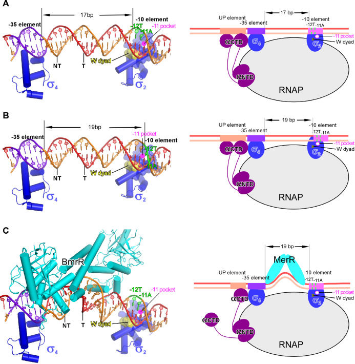
Transcription factors (TFs) modulate gene expression by regulating the accessibility of promoter DNA to RNA polymerases (RNAPs) in bacteria. The MerR family TFs are a large class of bacterial proteins unique in their physiological functions and molecular action: they function as transcription repressors under normal circumstances, but rapidly transform to transcription activators under various cellular triggers, including oxidative stress, imbalance of cellular metal ions, and antibiotic challenge. The promoters regulated by MerR TFs typically contain an abnormal long spacer between the -35 and -10 elements, where MerR TFs bind and regulate transcription activity through unique mechanisms. In this review, we summarize the function, ligand reception, DNA recognition, and molecular mechanism of transcription regulation of MerR-family TFs.

Keywords: MerR; RNA polymerase; gene expression; gene transcription; transcription factor.


Conflict of interest statement

The authors declare that they have no conflict of interest.

Figures

None
Figure1
Models for bacterial transcription initiation and canonical transcription activation(A) The model of RNAP-promoter DNA closed complex (RPc; upper panel) and RNAP-promoter DNA open complex (RPo; lower panel). In RPc, the double-stranded –35 element is recognized by σ4 in a sequence-specific manner, while the double-stranded –10 element is presented onto the surface of σ2 and restrained by sequence nonspecific electrostatic attraction interactions. In RPo, a ~13 bp transcription bubble is unwound and stabilized inside of RNAP. The base pair of –11 position is forced open by the W-dyad (W433 and W434 in E. coli RNAP σ70), and the adenine base of –11A of the non-template strand is recognized and secured in the –11 pocket. The other domains of σ factors are hidden for clarity. (B) The models for Class I (upper panel) and Class II (lower panel) transcription activation. A Class I transcription activator binds to the upstream of core promoter region and makes interactions with the C-terminal domain of the RNAP-α subunit (RNAP-αCTD). A Class II transcription activator binds to the proximal upstream of the core promoter region and makes interactions with both the RNAP-αCTD and σ4.
None
Figure2
The MerR-family TFs(A) The schematic of three categories of MerR-family TFs. DBD, DNA-binding domain; DH, dimerization helix; LBD, ligand-binding domain. (B) The structure of E. coli CueR dimer, a representative member of the metal-responsive MerR TFs, adapted from the crystal structure of Ag+-bound E. coli CueR-DNA complex (PDB: 4WLW). One protomer is colored in gray, and the second protomer is colored in orange (DBD), green (DH), and pink (LBD). The Ag+ is shows in gray sphere. (C) The structure of E. coli SoxR dimer, a representative member of the redox-responsive MerR TFs, adapted from the crystal structure of oxidated SoxR-DNA complex (PDB: 2ZHG). The [2Fe-2S] cluster is shown as sphere. (D) The structure of B. subtilis BmrR dimer, a representative member of the multidrug-resistance MerR TFs, adapted from the crystal structure of puromycin-bound BmrR-DNA complex (PDB: 1EXI). The puromycin is shown as sphere.
None
Figure3
DNA recognition of MerR-family TFs(A) The consensus DNA sequence logo of E. coli CueR regulated promoters. The palindromic repeats are highlighted by arrows. The positions are numbered respective to the transcription start site (+1). (B) The interaction between the CueR-DBD and dsDNA. The sequence nonspecific interactions between backbone phosphates of DNA and residues of CueR-DBD are shown in the middle panel. The base-specific interactions made by CueR-DBD are shown in the right panel. (C) The consensus protein sequence logo of CueR from various bacterial species. (D) The multiple-sequence alignment of DBD from multiple MerR TFs. The residues contacting backbone phosphates are labeled by diamonds and the residues making base-specific interactions are labeled by asterisks.
None
Figure4
The signal reception of MerR-family TFs(A) The multiple sequence alignment and consensus protein sequence logo for the ligand-binding domain of MerR-family TFs. The two mostly conserved cysteines are highlighted and labeled by asterisks. (B) E. coli CueR coordinates one molecule of Ag+ through Cys112 and Cys120 of its metal-binding loop. The Ser77’ of the other protomer restrains the conformation of Cys112 (PDB: 4WLW). (C) P. putida CadR coordinates one molecule of Cd+ through Cys112 and Cys119 from one protomer and Cys77’ from the other protomer (PDB: 6JGX). (D) B. megaterium MerR coordinates one molecule of Hg2+ through Cys117 and Cys126 from one protomer and Cys82’ from the other protomer (PDB: 5CRL). (E) E. coli ZntR coordinates two molecules of Zn+ through Cys114, Cys115, H119, and Cys124 from one protomer, Cys79’ from the other protomer, and a phosphate group (PDB: 1Q08). (F) E. coli SoxR coordinates the [2Fe-2S] cluster through residues Cys119, Cys 122, Cys 124, and Cys 130 of the same protomer. (G) Structure superimposition of the ligand-binding domains of four multidrug-resistance MerR TFs, B. subtilis BmrR (cyan; PDB: 3IAO), E. coli EcmrR (green; PDB: 6XLA), P. aeruginosa BrlR (blue; PDB: 5XBT), and E. coli SbmC (light brown; PDB: 1JYH; DNA Gyrase inhibitory protein, Gyrl). (H-J) Detailed presentation of the ligand-binding pocket of B. subtilis BmrR occupied by puromycin (PDB: 3Q3D), ethidium (PDB: 3Q2Y), and kanamycin (PDB: 3Q5R). Residues V147 and I255 server as a hydrophobic pincer. Residues Y152, Y170, Y187, F224, Y229, and Y268 server as an aromatic ring to accommodate drugs with distinct chemical structures. Residue E253 makes auxiliary polar interactions with the drugs.
None
Figure5
The cryo-EM structures of transcription activation complexes comprising MerR-family TFsThe cryo-EM structures of (A) E. coli CueR transcription activation complex (PDB: 6XH7), (B) E. coli EcmrR transcription activation complex (PDB: 6XL5), and (C) B. subtilis BmrR transcription activation complex (PDB: 7CKQ). The insertion box shows the small surface patch of CueR-DBD that interacts with the σNCR. (D) Structure superimposition of the upstream dsDNAs of the above three transcription activation complexes and that of a bacterial RPo (PDB: 6OUL). (E) The kinks of the upstream promoter DNA at positions −35 (⦟1, 28°), −30 (⦟2, 44°), −24 (⦟3, 85°) and −18 (⦟4, 40°) in the cryo-EM structure of E. coli CueR-TAC (PDB: 6LDI). Kink 1 at −35 is induced by CueR and σ704, and kinks 2, 3 and 4 are induced by the CueR dimer.
None
Figure6
The DNA-distortion mechanism of transcription activation by MerR-family TFs(A) The –35 and –10 elements are properly aligned on the protein surface of σ4 and σ2. The –11A of the nontemplate strand DNA is close to the –11 pocket. The panel figure is prepared from a B. subtilis RPc model comprising a promoter DNA with 17-bp –35/–10 spacer. (B) The –11A of the nontemplate strand DNA is rotated away from the –11 pocket in the B. subtilis RPc model comprising a promoter DNA with 19-bp –35/–10 spacer. (C) The BmrR-induced central kink realigns the −35 and −10 elements to a proper space and phase for simultaneous engagement by σ4 and σ2 according to the structure model of a BmrR-RPc complex. Yellow surfaces (left) and arrows (right) show the tryptophan dyad; pink circles show the –11 pocket.

Similar articles

Cited by

References

    1. Roeder RG, Rutter WJ. Multiple forms of DNA-dependent RNA polymerase in eukaryotic organisms. Nature. . 1969;224:234–237. doi: 10.1038/224234a0. - DOI - PubMed
    1. Chamberlin M, Berg P. Deoxyribo ucleic acid-directed synthesis of ribonucleic acid by an enzyme from Escherichia coli . Proc Natl Acad Sci U S A. . 1962;48:81–94. doi: 10.1073/pnas.48.1.81. - DOI - PMC - PubMed
    1. Feklistov A, Sharon BD, Darst SA, Gross CA. Bacterial sigma factors: a historical, structural, and genomic perspective. Annu Rev Microbiol. . 2014;68:357–376. doi: 10.1146/annurev-micro-092412-155737. - DOI - PubMed
    1. Seshasayee AS, Sivaraman K, Luscombe NM. An overview of prokaryotic transcription factors: a summary of function and occurrence in bacterial genomes. Subcell Biochem. 2011,52: 7-23 - PubMed
    1. Browning DF, Busby SJW. Local and global regulation of transcription initiation in bacteria. Nat Rev Microbiol. . 2016;14:638–650. doi: 10.1038/nrmicro.2016.103. - DOI - PubMed

MeSH terms