. 2022 Jan 25;54(2):187-198.
doi: 10.3724/abbs.2021017.

The residues 4 to 6 at the N-terminus in particular modulate fibril propagation of β-microglobulin

Affiliations

The residues 4 to 6 at the N-terminus in particular modulate fibril propagation of β-microglobulin

Haibin Dang et al. Acta Biochim Biophys Sin (Shanghai). .

Abstract

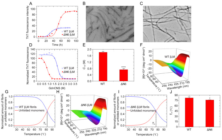
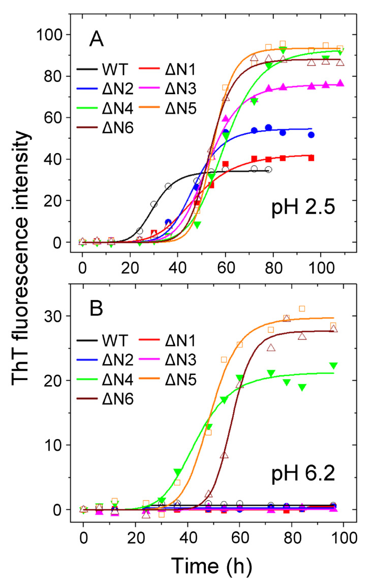
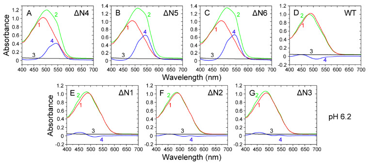
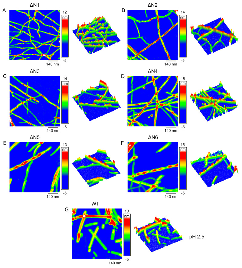
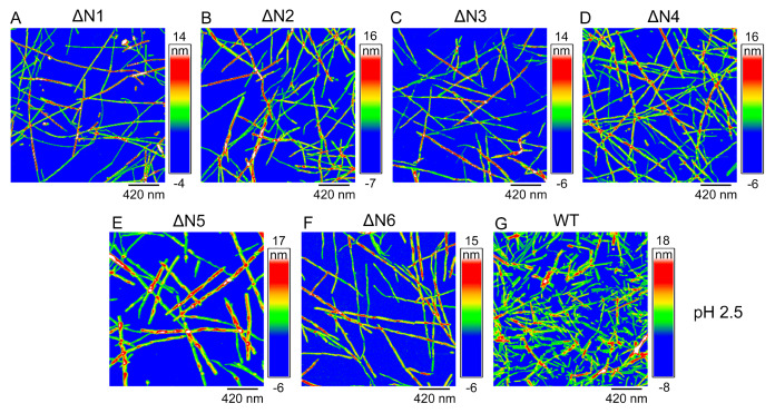
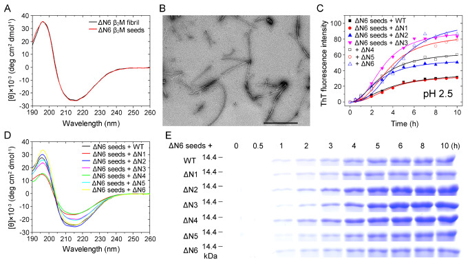
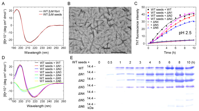
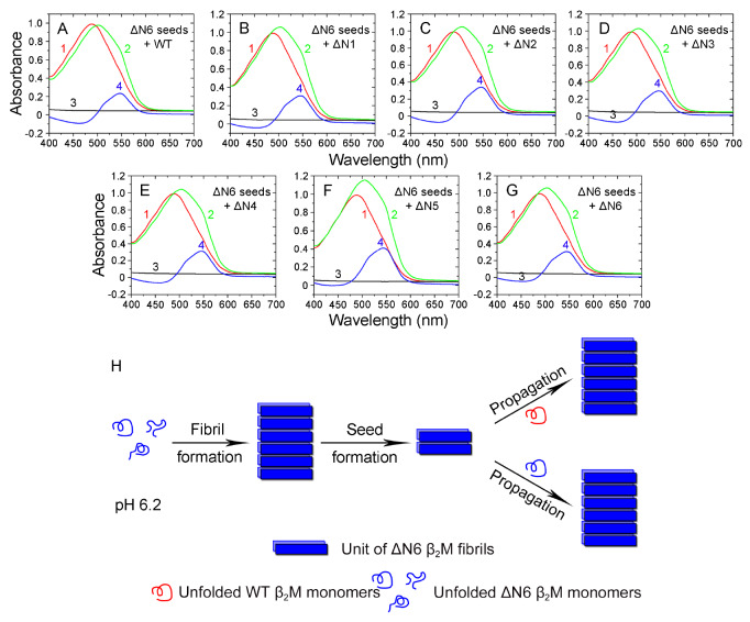
The ΔN6 truncation is the main posttranslational modification of β-microglobulin (βM) found in dialysis-related amyloid. Investigation of the interaction of wild-type (WT) βM with N-terminally truncated variants is therefore of medical relevance. However, it is unclear which residues among the six residues at the N-terminus are crucial to the interactions and the modulation of amyloid fibril propagation of βM. We herein analyzed homo- and heterotypic seeding of amyloid fibrils of WT human βM and its N-terminally-truncated variants ΔN1 to ΔN6, lacking up to six residues at the N-terminus. At acidic pH 2.5, we produced amyloid fibrils from recombinant, WT βM and its six truncated variants, and found that ΔN6 βM fibrils exhibit a significantly lower conformational stability than WT βM fibrils. Importantly, under more physiological conditions (pH 6.2), we assembled amyloid fibrils only from recombinant, ΔN4, ΔN5, and ΔN6 βM but not from WT βM and its three truncated variants ΔN1 to ΔN3. Notably, the removal of the six, five or four residues at the N-terminus leads to enhanced fibril formation, and homo- and heterotypic seeding of ΔN6 fibrils strongly promotes amyloid fibril formation of WT βM and its six truncated variants, including at more physiological pH 6.2. Collectively, these results demonstrated that the residues 4 to 6 at the N-terminus particularly modulate amyloid fibril propagation of βM and the interactions of WT βM with N-terminally truncated variants, potentially indicating the direct relevance to the involvement of the protein's aggregation in dialysis-related amyloidosis.

Keywords: amyloid fibril; atomic force microscopy; conformational stability; fibril propagation; human β-microglobulin; truncated variant.


Conflict of interest statement

The authors declare that they have no conflict of interest.

Figures

None
Figure1
ΔN6 β 2M fibrils exhibite a significantly lower conformational stability than WT β 2M fibrils at acidic pH 2.5 (A) Samples (85 μM) of WT β2M (blue) and ΔN6 β2M (red) were incubated at pH 2.5 with agitation at 220 rpm and 37°C. The solid lines show the best sigmoidal fit for the ThT intensity-time curves. (B, C) TEM images of amyloid fibrils formed by WT β2M (B) incubated for 84 h and ΔN6 β2M (C) incubated for 102 h at pH 2.5. Scale bar: 500 nm. (D) GdnCNS concentration-dependent denaturation profiles monitored by ThT fluorescence for amyloid fibrils produced from WT β2M (blue) and ΔN6 β2M (red) at pH 2.5, which were incubated for 1 h at 25°C with increasing concentrations of GdnCNS. (E) The C1/2 values for amyloid fibrils of WT β2M and ΔN6 β2M were determined using a sigmoidal equation and are expressed as the mean±SD of the values obtained from 3 independent experiments. C1/2, P = 0.038. (F-I) Three-dimensional (3D) diagrams of the CD spectra against temperature ranged from 25°C to 95°C for amyloid fibrils (25 μM) of WT β2M (F) and ΔN6 β2M (H) formed at pH 2.5; the normalized amount of the fibrils (blue) and the unfolded monomers (red) for WT β2M (G) and ΔN6 β2M (I). The isosbestic points represent the corresponding Tm values. (J) The Tm values were also determined using a sigmoidal equation and are expressed as the mean±SD of the values obtained from 3 independent experiments. Tm, P= 0.000021. The Student’s t test was used to perform statistical analyses. Values of P<0.05 indicate statistically significant differences. *P<0.05, and ****P<0.0001 relative to WT β2M (a control).
None
Figure2
Modulation of amyloid fibril formation of β 2M by residues 4 to 6 at the N-terminus under more physiological conditions (pH 6.2) analyzed by ThT binding assays (A,B) Amyloid fibril formation of WT β2M and its six truncated variants at pH 6.2 or 2.5. Samples (85 μM) of WT β2M (black circle) and its truncated mutants ΔN1 (red square), ΔN2 (blue circle), ΔN3 (magenta triangle), ΔN4 (green inverse triangle), ΔN5 (orange square), and ΔN6 (wine triangle) were incubated at either pH 2.5 (A) or pH 6.2 (B) with agitation at 220 rpm and 37°C. The solid lines show the best sigmoidal fit for the ThT intensity-time curves. At acidic pH 2.5, we produced amyloid fibrils from WT β2M and its six truncated variants. At more physiological pH 6.2, however, we assembled amyloid fibrils only from ΔN4, ΔN5, and ΔN6 β2M but not from WT β2M and its three truncated variants ΔN1 to ΔN3. All ThT binding assays were repeated at least three times, and the results were reproducible.
None
Figure3
Modulation of amyloid fibril formation of β 2M by residues 4 to 6 at the N-terminus under more physiological conditions (pH 6.2) analyzed by Congo red binding assays Amyloid fibrils of WT β2M and its N-terminally-truncated variants were formed at pH 6.2 with agitation at 220 rpm and 37°C. Absorbance data are shown for amyloid fibrils at the end of fibril formation for 10 μM WT β2M (D) and its truncated mutants ΔN1 (E), ΔN2 (F), ΔN3 (G), ΔN4 (A), ΔN5 (B), and ΔN6 (C) in the presence of 50 μM Congo red at 25°C. The difference spectra (Curve 4, blue) with the maximum absorbance at 550 nm were obtained by subtracting the absorbance spectra of β2M fibrils alone (Curve 3, black) and Congo red alone (Curve 1, red) with the maximum absorbance at 490 nm from those of β2M fibrils + Congo red (Curve 2, green). At pH 6.2, we assembled amyloid fibrils only from ΔN4, ΔN5, and ΔN6 β2M but not from WT β2M and its three truncated variants ΔN1 to ΔN3. All Congo red binding assays were repeated at least three times, and the results were reproducible.
None
Figure5
High-resolution 3D AFM images of β 2M fibrils at acidic pH 2.5 2D (left) and 3D (right) AFM images of amyloid fibrils formed by WT β2M incubated for 78 h (A), ΔN1 (B) and ΔN2 (C) incubated for 96 h, and ΔN3 (D), ΔN4 (E), ΔN5 (F), and ΔN6 (G) incubated for 108 h at pH 2.5 with agitation at 220 rpm and 37°C. Scale bar: 140 nm.
None
Figure4
High-resolution AFM images of β 2M fibrils at acidic pH 2.5 AFM images of amyloid fibrils formed by WT β2M incubated for 78 h (G), ΔN1 (A), and ΔN2 (B) incubated for 96 h, and ΔN3 (C), ΔN4 (D), ΔN5 (E), and ΔN6 (F) incubated for 108 h at pH 2.5 with agitation at 220 rpm and 37°C. The three different time points (78, 96, and 108 h) were used because the incubation times at the end of fibril formation for WT β2M and ΔN1 to ΔN6 were different. Scale bar: 420 nm. At pH 2.5, we produced amyloid fibrils from WT β2M and its six truncated variants.
None
Figure6
Homo- and heterotypic seeding of ΔN6 fibrils strongly promoted amyloid fibril formation of WT β 2M and its six truncated variants at acidic pH 2.5 (A) The secondary structures of ΔN6 fibrils (black) and ΔN6 fibril seeds (red) at pH 2.5 monitored by far-UV CD. (B) TEM images of ΔN6 fibril seeds at pH 2.5. Scale bar: 500 nm. (C) Samples (85 μM) of WT β2M (solid square) and its six truncated mutants ΔN1 (solid circle), ΔN2 (solid up triangle), ΔN3 (solid inverse triangle), ΔN4 (open square), ΔN5 (open circle), and ΔN6 (open triangle) in the presence of 2% (v/v) ΔN6 fibril seeds were incubated at pH 2.5 with agitation at 220 rpm and 37°C. The solid lines show the best fit for the ThT intensity-time curves. (D) CD spectra at the end of fibril formation (10 h) for WT β2M (black) and its six truncated variants ΔN1 (red), ΔN2 (blue), ΔN3 (magenta), ΔN4 (green), ΔN5 (cyan), and ΔN6 (yellow). (E) SDS-PAGE analysis of time-dependent sarkosyl-insoluble β2M incubated with 2% (v/v) ΔN6 fibril seeds at pH 2.5, including WT β2M and its six truncated mutants. 85 μM β2M samples were incubated with 2% sarkosyl and separated by 15% SDS-PAGE. The insoluble β2M monomers were detected by Coomassie Blue R250 staining.
None
Figure7
Homo- and heterotypic seeding of WT β 2M fibrils facilitated amyloid fibril formation of the wild-type protein and its three truncated variants ΔN1 to ΔN3 but not ΔN4, ΔN5, and ΔN6 at acidic pH 2.5 (A) The secondary structures of WT β2M fibrils (black) and WT β2M fibril seeds (red) at pH 2.5 monitored by far-UV CD. (B) TEM images of WT β2M fibril seeds at pH 2.5. Scale bar: 500 nm. (C) Samples (85 μM) of WT β2M (solid square) and its six truncated mutants ΔN1 (solid circle), ΔN2 (solid up triangle), ΔN3 (solid inverse triangle), ΔN4 (open square), ΔN5 (open circle), and ΔN6 (open triangle) in the presence of 2% (v/v) WT β2M fibril seeds were incubated at pH 2.5 with agitation at 220 rpm and 37°C. The solid lines show the best fit for the ThT intensity-time curves. (D) CD spectra at the end of fibril formation (10 h) for WT β2M (black) and its six truncated variants ΔN1 (red), ΔN2 (blue), ΔN3 (magenta), ΔN4 (green), ΔN5 (cyan), and ΔN6 (yellow). (E) SDS-PAGE analysis of time-dependent sarkosyl-insoluble β2M incubated with 2% (v/v) WT β2M fibril seeds at pH 2.5, including WT β2M and its six truncated mutants.
None
Figure8
Homo- and heterotypic seeding of ΔN6 fibrils strongly promoted amyloid fibril formation of WT β 2M and its six truncated variants at more physiological conditions (pH 6.2) (A–G) Amyloid fibrils of WT β2M and its N-terminally-truncated variants were induced by 10% (v/v) ΔN6 fibril seeds at pH 6.2 with agitation at 220 rpm and 37°C. Absorbance data are shown for amyloid fibrils at the end of fibril formation for 10 μM WT β2M (A) and its truncated mutants ΔN1 (B), ΔN2 (C), ΔN3 (D), ΔN4 (E), ΔN5 (F), and ΔN6 (G) in the presence of 50 μM Congo red at 25°C. The difference spectra (Curve 4, blue) with the maximum absorbance at 550 nm were obtained by subtracting the absorbance spectra of β2M fibrils alone (Curve 3, black) and Congo red alone (Curve 1, red) with the maximum absorbance at 490 nm from those of β2M fibrils + Congo red (Curve 2, green). All Congo red binding assays were repeated at least three times, and the results were reproducible. (H) A hypothetical model showing how the six residues at the N-terminus modulate fibril propagation of β2M and the interactions of WT β2M with N-terminally truncated variants at more physiological pH 6.2. Under such conditions, the loss of the first six residues (ΔN6) facilitates amyloid fibril (blue brick) formation from ΔN6 β2M monomers (blue rope), and homo- and heterotypic seeding of ΔN6 fibrils strongly promotes amyloid fibril formation of WT β2M (red rope) and its truncated variant ΔN6 (blue rope).

Similar articles

References

    1. Dobson CM. Protein folding and misfolding. Nature. . 2003;426:884–890. doi: 10.1038/nature02261. - DOI - PubMed
    1. Chiti F, Dobson CM. Protein misfolding, functional amyloid, and human disease. Annu Rev Biochem. . 2006;75:333–366. doi: 10.1146/annurev.biochem.75.101304.123901. - DOI - PubMed
    1. Goedert M, Spillantini MG. A century of Alzheimer’s disease. Science. . 2006;314:777–781. doi: 10.1126/science.1132814. - DOI - PubMed
    1. Spillantini MG, Luise Schmidt M, Lee VM, Trojanowski JQ, Jakes R, Goedert M. α-Synuclein in Lewy bodies. Nature. . 1997;388:839–840. doi: 10.1038/42166. - DOI - PubMed
    1. Owen MC, Gnutt D, Gao M, Wärmländer SK, Jarvet J, Gräslund A, Winter R, et al. Effects of in vivo conditions on amyloid aggregation . Chem Soc Rev. . 2019;48:3946–3996. doi: 10.1039/C8CS00034D. - DOI - PubMed