. 2021 Feb 22;13(1):e0356121.
doi: 10.1128/mbio.03561-21. Epub 2022 Feb 8.

Novel CRK-Cyclin Complex Controls Spindle Assembly Checkpoint in Toxoplasma Endodyogeny

Affiliations

Novel CRK-Cyclin Complex Controls Spindle Assembly Checkpoint in Toxoplasma Endodyogeny

Lauren M Hawkins et al. mBio. .

Abstract

Opportunistic parasites of the Apicomplexa phylum use a variety of division modes built on two types of cell cycles that incorporate two distinctive mechanisms of mitosis: uncoupled from and coupled to parasite budding. Parasites have evolved novel factors to regulate such unique replication mechanisms that are poorly understood. Here, we have combined genetics, quantitative fluorescence microscopy, and global proteomics approaches to examine endodyogeny in Toxoplasma gondii dividing by mitosis coupled to cytokinesis. In the current study, we focus on the steps controlled by the recently described atypical Cdk-related kinase T. gondii Crk6 (TgCrk6). While inspecting protein complexes, we found that this previously orphaned TgCrk6 kinase interacts with a parasite-specific atypical cyclin, TgCyc1. We built conditional expression models and examined primary cell cycle defects caused by the lack of TgCrk6 or TgCyc1. Quantitative microscopy assays revealed that tachyzoites deficient in either TgCrk6 or the cyclin partner TgCyc1 exhibit identical mitotic defects, suggesting cooperative action of the complex components. Further examination of the mitotic structures indicated that the TgCrk6/TgCyc1 complex regulates metaphase. This novel finding confirms a functional spindle assembly checkpoint (SAC) in T. gondii. Measuring global changes in protein expression and phosphorylation, we found evidence that canonical activities of the Toxoplasma SAC are intertwined with parasite-specific tasks. Analysis of phosphorylation motifs suggests that Toxoplasma metaphase is regulated by CDK, mitogen-activated kinase (MAPK), and Aurora kinases, while the TgCrk6/TgCyc1 complex specifically controls the centromere-associated network. IMPORTANCE The rate of Toxoplasma tachyzoite division directly correlates with the severity of the disease, toxoplasmosis, which affects humans and animals. Thus, a better understanding of the tachyzoite cell cycle would offer much-needed efficient tools to control the acute stage of infection. Although tachyzoites divide by binary division, the cell cycle architecture and regulation differ significantly from the conventional binary fission of their host cells. Unlike the unidirectional conventional cell cycle, the Toxoplasma budding cycle is braided and is regulated by multiple essential Cdk-related kinases (Crks) that emerged in the place of missing conventional cell cycle regulators. How these novel Crks control apicomplexan cell cycles is largely unknown. Here, we have discovered a novel parasite-specific complex, TgCrk6/TgCyc1, that orchestrates a major mitotic event, the spindle assembly checkpoint. We demonstrated that tachyzoites incorporated parasite-specific tasks in the canonical checkpoint functions.

Keywords: Apicomplexa; Toxoplasma gondii; apicomplexan parasites; cell cycle; cyclin; cyclin-dependent kinase; endodyogeny; mitosis; protein phosphorylation; spindle assembly checkpoint.


Conflict of interest statement

The authors declare no conflict of interest.

Figures

FIG 1
FIG 1
Characterization of the TgCrk6 in the new conditional expression model. (A) Diagram shows the mechanism of auxin-induced degradation of AID-tagged TgCrk6. In the absence of plant hormone auxin (IAA), the native promoter controls TgCrk6AID-HA expression at endogenous levels. Addition of 500 μM auxin (yellow star) promotes interaction of AID-modified TgCrk6 with F-box protein TIR1 (dark blue) and ubiquitin ligase SCF (light blue), resulting in the rapid degradation of TgCrk6AID-HA by proteasome. (B) The differential interference contrast (DIC) images of the HFF monolayers infected with RH TgCrk6AID-HA tachyzoites and grown with or without 500 μM auxin for 7 days. Note that only TgCrk6-expressing tachyzoites (−auxin) formed viable plaques. (C) Western blot analysis of the total lysates of the RHΔKu80TIR1 tachyzoites expressing TgCrk6AID-HA. Lysates of nontreated parasites and parasites treated with auxin for 10 min and chased for 1 h were analyzed. Western blots were probed with α-HA (α-rat IgG-HRP) to detect TgCrk6 and with α-tubulin A (α-mouse IgG-HRP) to confirm equal loading of the total lysates. MW, molecular weight. (D) Schematics of the tachyzoite cell cycle. The circle represents 7 h of the tachyzoite division at 37°C and indicates the relative position of the cell cycle phases. Drawings show morphological changes of the dividing tachyzoite. Red color represents the time and primary localization of the TgCrk6 kinase deduced from the quantitative immunofluorescent analysis (see details below). Purple, centrosome (marker, Centrin1); green, centrocone and basal complex (marker, MORN1); blue, nucleus (marker, 4′,6-diamidino-2-phenylindole [DAPI]); black, surface alveoli (marker, IMC1). (E) IF microscopy analysis of TgCrk6AID-HA expression in RHΔKu80TIR1 tachyzoites. To reveal cell cycle-dependent expression, TgCrk6AID-HA (α-HA/α-rat IgG Fluor 568) was costained with centrosomes (α-Centrin1/α-mouse IgG Fluor 488), centrocone (α-MORN1/α-rabbit IgG Flour 488), or alveolar protein IMC1 (α-IMC1/α-rabbit IgG Fluor 488). Cell cycle phases were determined based on the number and morphology of the reference structures, indicated with arrows. Double-headed arrows point to TgCrk6 accumulated in the centrocone. The percentage of the parasites with single or duplicated centrosomes and with internal buds, expressing or not expressing TgCrk6AID-HA, was calculated from 3 independent experiments. Mean values ± standard deviations (SD) are shown under corresponding images. Scale bar, 5 μm.
FIG 2
FIG 2
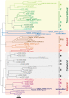
Phylogenetic analysis of apicomplexan cyclins. Phylogenetic tree based on multiple-sequence alignment of predicted cyclins from Toxoplasma gondii, Plasmodium falciparum, Chromera velia, Cryptosporidium parvum, Babesia bovis, Homo sapiens, and Mus musculus. The topology of the phylogenetic tree is optimized by maximum likelihood. Topology support from bootstrapping is shown at nodes. Three classes of cyclins are indicated with different colors. Boldfaced lettering highlights five new T. gondii cyclins.
FIG 3
FIG 3
TgCrk6 interacts with novel mitotic cyclin TgCyc1. (A) Selected TgCrk6 interactors identified by mass spectroscopy analysis of isolated TgCrk6 complexes. The table depicts selected hits predicted to localize to the nucleus. FC, fold change of the peptides in the TgCrk6 complexes. IP, immunoprecipitation. (B) TgCyc1 forms a complex with TgCrk6. TgCyc1/TgCrk6 complexes were immunoisolated from the soluble fraction (In; input) of parasites coexpressing endogenous TgCyc1AID-HA and TgCrk6myc. Beads with precipitated complexes (B) and depleted soluble fraction (Un) (unbound) were probed with α-myc (α-rabbit IgG-HRP) and α-HA (α-rat IgG-HRP) antibodies to detect interacting TgCrk6 and to confirm efficient pulldown of TgCyc1 (the IP panels at the top). (C) IFA images of tachyzoites coexpressing endogenous TgCyc1AID-HA and TgCrk6myc. Parasites were costained with α-HA/α-rat IgG Fluor 568 and α-myc/α-rabbit IgG Fluor 488. The enlarged overlay image shows an example of colocalization (black arrow) and separate localization, indicated with the corresponding color of the arrows. Scale bar, 5 μm. (D) Selected nuclear TgCrk6 interactors from panel A are maximally expressed in S phase and mitosis. Heatmap shows transcriptional signatures of selected genes across tachyzoite cycle of T. gondii. The transcriptional profile of each gene is normalized by z score. (E) Western blot analysis of the total lysates of the RHΔKu80TIR1 tachyzoites expressing TgCyc1AID-HA. Lysates of nontreated parasites and parasites treated with auxin for 10 min and chased for 1 h were analyzed. Western blots were probed with α-HA (α-rat IgG-HRP) to detect TgCyc1 and with α-tubulin A (α-mouse IgG-HRP) to confirm equal loading of the total lysates. (F) The new cyclin essentiality was tested by the ability of RH TgCyc1AID-HA mutant parasites to form plaques after 7 days with or without 500 μM auxin. The representative DIC images are shown. Note that only TgCyc1-expressing tachyzoites (−auxin) formed viable plaques. (G) IF microscopy analysis of TgCyc1AID-HA expression in RHΔKu80TIR1 tachyzoites. To establish the time frame for TgCyc1 expression, TgCyc1AID-HA (α-HA/α-rat IgG Fluor 568) was costained with centrosomes (α-Centrin1/α-mouse IgG Fluor 488), centrocone (α-MORN1/α-rabbit IgG Flour 488), or alveolar protein IMC1 (α-IMC1/α-rabbit IgG Fluor 488). Cell cycle phases were determined based on the number and morphology of the reference structures, indicated with arrows. Double-headed arrows point to TgCyc1 accumulated in the centrocone. The percentage of the parasites with a single or duplicated centrosome and with internal buds, expressing or not expressing TgCyc1AID-HA, was calculated from 3 independent experiments. Mean value ±SD is shown under corresponding images. Scale bar, 5 μm. (H) Schematics of the tachyzoite cell cycle. The circle represents 7 h of the tachyzoite division at 37°C and indicates the relative position of the cell cycle phases. Drawings show morphological changes of the dividing tachyzoite. Red color represents the time and primary localization of the novel cyclin TgCyc1, deduced from the quantitative immunofluorescent analysis (see details for panel G). Purple, centrosome (marker, Centrin1); green, centrocone and basal complex (marker, MORN1); blue, nucleus (marker, DAPI); black, surface alveoli (marker, IMC1).
FIG 4
FIG 4
Lack of TgCrk6 or TgCyc1 leads to similar arrest in mitosis. (A) The RH TgCrk6AID-HA (upper graph) or RH TgCyc1AID-HA (lower graph) tachyzoites were examined by IFA after 3, 4, 5, 6, and 7 h with 500 μM auxin and compared to the untreated population (0 h). Centrosome duplication and budding were evaluated using α-Centrin1 (red bars) and α-IMC1 (green bars) antibody. Parasites were costained with DAPI to detect nuclear morphology. All markers were accounted for to quantify abnormalities of the cell division (blue dotted line): DNA missegregation and centrosome overduplication. Each cell cycle marker was examined in 100 random vacuoles in three independent experiments. The mean and SD values are show on the plots. (B) The IFA images depict cellular abnormalities accumulated after 7 h of TgCyc1 depletion (7 h, auxin). Parasite, nuclear morphology, and centrosome numbers were detected with α-IMC1/α-rabbit IgG fluor 488, DAPI (blue), and α-Centrin1/α-mouse IgG Fluor 568 antibodies and stains. Scale bar, 5 μm. (C) The reversibility of the mitotic block induced by TgCrk6 or TgCyc1 withdrawal was determined by plaque assay. Freshly invaded RH TgCrk6AID-HA (blue line) or RH TgCyc1AID-HA (red line) parasites were incubated with 500 μM auxin for the indicated times before the medium was replaced with normal growth medium without auxin to allow for plaque development. Plaque numbers represent averages from three independent measurements. (D) Quantification of the primary mitotic defect caused by RH TgCrk6AID-HA or RH TgCyc1AID-HA deficiency. Unresolved (orange bars) and resolved (red bars) centrocones were quantified based on the MORN1 and IMC1 costaining shown in panel B. One hundred random vacuoles of the parasites containing a single centrocone dot with two attached rings (metaphase) or two centrocone dots with an attached ring (anaphase and early budding) were examined in three independent experiments. The mean values are plotted on the graph.
FIG 5
FIG 5
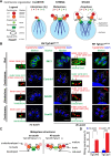
TgCrk6/TgCyc1 complex controls metaphase-to-anaphase transition. (A) Schematics of the interphase, metaphase, and anaphase nucleus and perinuclear structures of the Toxoplasma tachyzoite. Two cores of bipartite centrosome expand, duplicate, and separate, forming a distinctive pattern: a cluster in interphase, a string in metaphase, and a stack in anaphase. Present throughout the cell cycle, kinetochore and centromere segregate in anaphase of mitosis, changing the stoichiometry of the mitotic structures (colored numbers). The key mitotic structures and corresponding markers are listed in the legend on the right. (B) IFA analysis of the metaphase in the tachyzoites expressing or lacking TgCrk6 or TgCyc1. To visualize mitotic structures, tachyzoites expressing TgCEP250Myc (α-myc/α-rat IgG Fluor 568, inner and outer core of centrosome marker) were costained with organelle markers using the following antibodies: α-CenH3/α-mouse IgG Fluor 488 (centromere), α-Ndc80/α-mouse IgG Fluor 488 (kinetochore), α-Centrin1/α-mouse IgG Fluor 488 (outer centrosome), α-IMC1/α-mouse IgG Fluor 488 (bud), and DNA stain DAPI. The left panel shows undisturbed metaphase organization (no auxin). Two panels on the right show changes invoked by degradation of TgCrk6 or TgCyc1. Enlarged images on the side demonstrate relative positions of the visualized organelles (red and green channel only). Scale bar, 5 μm. (C) Summary of the IFA analysis in panel B. Schematics show metaphase structures of the parasites expressing (no auxin) and lacking (4 h auxin) TgCrk6/TgCyc1 complex. (D) Quantification of the string centrosome organization in tachyzoites expressing or deficient in TgCrk6 or TgCyc1. Centrosomes were examined in 100 random vacuoles in three independent experiments. The mean and SD values are show on the plots.
FIG 6
FIG 6
Spindle assembly checkpoint in Toxoplasma is regulated by phosphorylation and proteolysis. (A and B) Volcano plots show changes of the protein expression level (upper graph) and of the phosphosite intensity (lower graph) at the checkpoint block induced by TgCyc1 downregulation after 4 h. GO term enrichment analysis was performed on upregulated (orange) and downregulated (blue) groups. Results are shown on the sides of each plot. The bubbles reflect the size of individual pools. (C) The heat map displays global changes of the protein phosphorylation caused by auxin-induced TgCyc1 degradation for 30 min and 4 h. The proteins are organized according to the similarity in phosphorylation by K-means and combined into four classes based on the temporal dynamic of phosphorylation. (D) The phosphorylation motifs affected by 30 min of TgCyc1 degradation were deduced using MoMo software. Three dominant motifs and corresponding scores are shown. The responsible kinase family is indicated on the corresponding plot. (E) Table of the putative TgCrk6 substrates. The list was created based on the reduction of phosphorylation intensity within a proline-driven motif caused by the lack of TgCrk6 and TgCyc1 for 30 min. Affected phosphoserine residue is shown in red.

References

    1. Francia ME, Striepen B. 2014. Cell division in apicomplexan parasites. Nat Rev Microbiol 12:125–136. doi:10.1038/nrmicro3184. - DOI - PubMed
    1. Gubbels M-J, Coppens I, Zarringhalam K, Duraisingh MT, Engelberg K. 2021. The modular circuitry of apicomplexan cell division plasticity. Front Cell Infect Microbiol 11:670049. doi:10.3389/fcimb.2021.670049. - DOI - PMC - PubMed
    1. Gubbels M-J, Keroack CD, Dangoudoubiyam S, Worliczek HL, Paul AS, Bauwens C, Elsworth B, Engelberg K, Howe DK, Coppens I, Duraisingh MT. 2020. Fussing about fission: defining variety among mainstream and exotic apicomplexan cell division modes. Front Cell Infect Microbiol 10:269. doi:10.3389/fcimb.2020.00269. - DOI - PMC - PubMed
    1. White MW, Suvorova ES. 2018. Apicomplexa cell cycles: something old, borrowed, lost, and new. Trends Parasitol 34:759–771. doi:10.1016/j.pt.2018.07.006. - DOI - PMC - PubMed
    1. Brooks CF, Francia ME, Gissot M, Croken MM, Kim K, Striepen B. 2011. Toxoplasma gondii sequesters centromeres to a specific nuclear region throughout the cell cycle. Proc Natl Acad Sci USA 108:3767–3772. doi:10.1073/pnas.1006741108. - DOI - PMC - PubMed

LinkOut - more resources