. 2022 Dec;27(1):32-44.
doi: 10.1080/13510002.2022.2036507.

Lycium Barbarum polysaccharide protects HaCaT cells from PM2.5-induced apoptosis via inhibiting oxidative stress, ER stress and autophagy

Affiliations

Lycium Barbarum polysaccharide protects HaCaT cells from PM2.5-induced apoptosis via inhibiting oxidative stress, ER stress and autophagy

Sen Zhu et al. Redox Rep. 2022 Dec.

Abstract

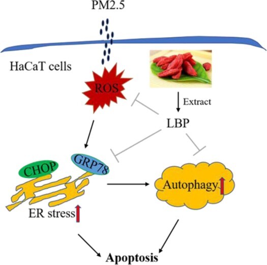
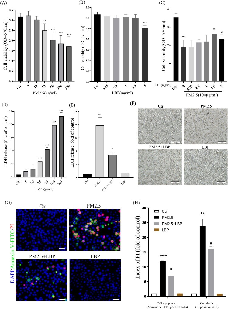
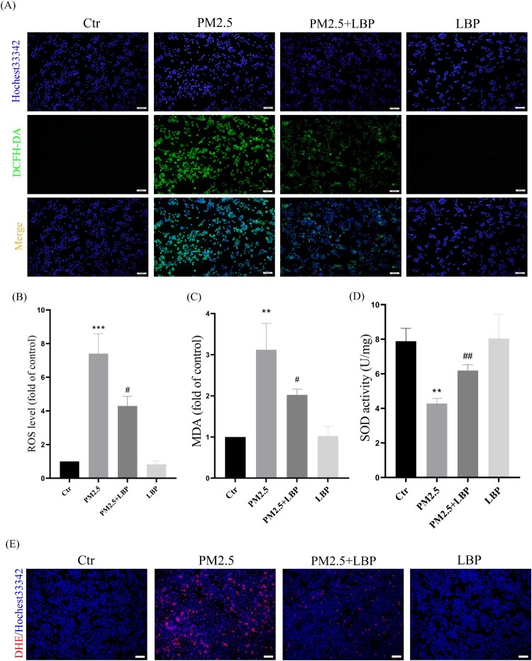
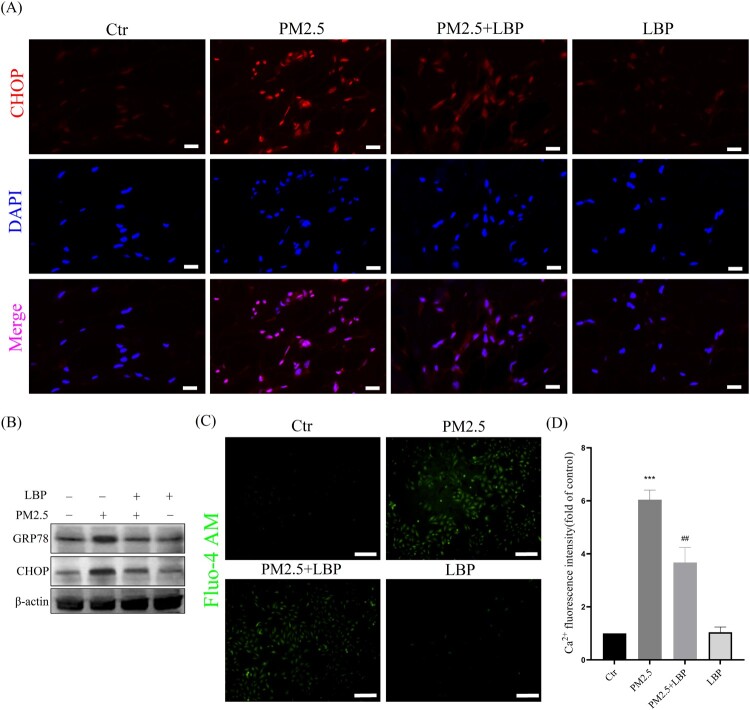
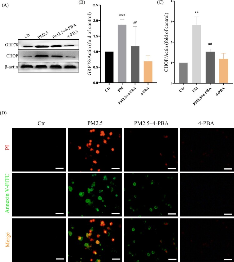
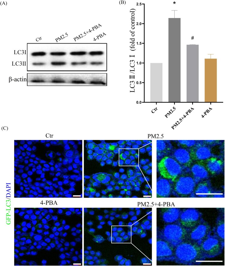
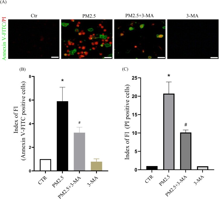
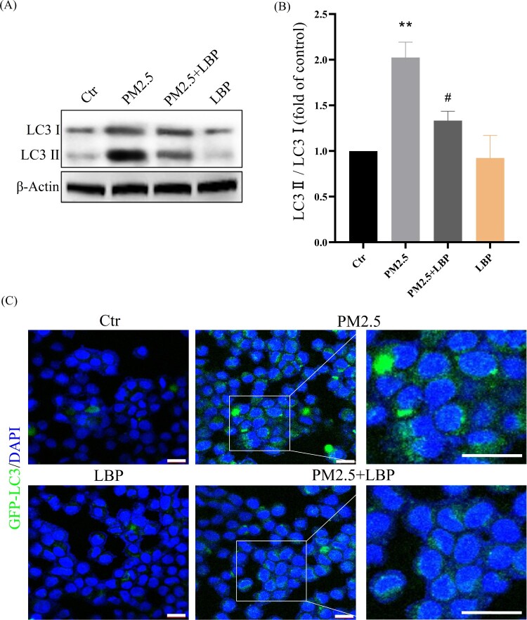
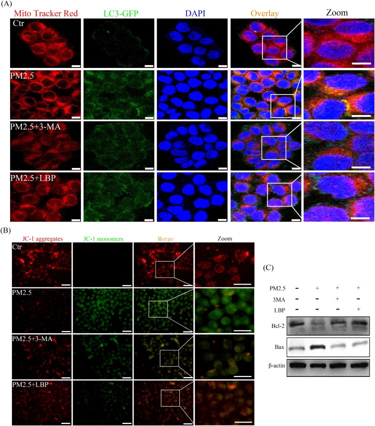
Objectives: Lycium barbarum polysaccharide (LBP) is a natural polysaccharide extracted from Lycium barbarum that has anti-inflammatory, anti-apoptotic and anti-aging effects, and plays a role in the prevention and treatment of various diseases. In this study, we investigated the therapeutic effect of LBP on particulate matter 2.5 (PM2.5)-induced skin damage.Methods: Cell viability was analyzed by MTT and LDH assays. Apoptosis was analyzed by Annexin V-FITC/PI staining. Oxidative stress/damage were assessed by intracellular ROS levels, MDA content and SOD activity. The intracellular protein expression was analyzed by Western blot. Mitochondrial damage was assayed by mitochondrial membrane potential with JC-1 probe. LC3-GFP adenovirus was transfected into HaCaT cells to analyze intracellular autophagosome levels.Results: In PM2.5-treated HaCaT cells, LBP pretreatment reduced PM2.5-induced cytotoxicity, ameliorated cell morphology and reduced cell apoptosis. LBP also inhibited the expression levels of GRP78 and CHOP, reduced the conversion of LC3I to LC3II, inhibited Bax protein and activated Bcl-2 protein. Furthermore, LBP inhibited PM2.5-induced mitochondrial autophagy (mitophagy) and mitochondrial damage. PM2.5-induced autophagy was regulated by endoplasmic reticulum (ER) stress.Conclusion: LBP protects skin cells from PM2.5-induced cytotoxicity by regulating the oxidative stress-ER stress-autophagy-apoptosis signaling axis, revealing that LBP has a great potential for the skin protection.

Keywords: Lycium barbarum polysaccharide (LBP); PM2.5; antioxidant; apoptosis; autophagy; endoplasmic reticulum (ER) stress; mitochondrial damage; oxidative damage.


Conflict of interest statement

No potential conflict of interest was reported by the author(s).

Figures

None
Graphical abstract
Figure 1.
Figure 1.
LBP reduces PM2.5-triggered apoptosis and increases cell survival. (A) MTT assay was used to detect the cytotoxicity of PM2.5 in HaCaT cells. (B) MTT assay was used to detect the effects of LBP on cell survival. (C) Cell viability was analyzed by MTT assay. HaCaT cells were pretreated with different concentrations of LBP and then co-incubated with PM2.5 for 24 h. (D) LDH assay was used to detect PM2.5-induced cytotoxicity in HaCaT cells. (E) Cell cytotoxicity was analyzed by LDH assay. HaCaT cells were pretreated with 2.5 mg/ml LBP and then co-incubated with PM2.5 for 24 h. (F) Cell morphology was analyzed by an inverted fluorescent microscope. Scale bar = 100 μm. (G) Apoptosis was analyzed with Annexin V-FITC, PI and DAPI staining, and observed under a fluorescent microscope. Scale bar = 50 μm. (H) Statistics of Annexin V-FITC and PI positive cells. Values are mean ± SD. *p < 0.05, **p < 0. 01, ***p < 0.001 versus the control group; #p < 0.05, ##p < 0.01, ###p < 0.001 versus the PM2.5 treatment alone group.
Figure 2.
Figure 2.
LBP alleviates PM2.5-induced oxidative stress and oxidative damage in HaCaT cells. (A) Cells were double-stained with Hochest33342 and DCFH-DA, and intracellular ROS levels were observed by a fluorescent microscopy. Scale bar = 100 μm. (B) Statistics of intracellular ROS level, stained with DCFH-DA. The intracellular MDA level (C) and SOD activity (D) were detected by the corresponding assay kits. (E) Cells were double-stained with Hochest33342 and DHE, and intracellular ROS levels were observed by a fluorescent microscopy. Scale bar = 100 μm. Values are mean ± SD. *p < 0.05, **p < 0. 01, ***p < 0.001 versus the control group; #p < 0.05, ##p < 0.01, ###p < 0.001 versus the PM2.5 treatment alone group.
Figure 3.
Figure 3.
LBP inhibits PM2.5-induced ER stress and CHOP expression in HaCaT cells. (A) Immunofluorescence was used to detect the expression of CHOP in HaCaT cells. Scale bar = 20 μm. (B) Western blot assay was used to detect the protein levels of GRP78 and CHOP. (C) Intracellular Ca2+ was labeled with Fluo-4 AM and analyzed by a fluorescent microscope. Scale bar = 100 μm. (D) Statistics of intracellular Ca2+ level. Values are mean ± SD. *p < 0. 05 versus the control group; # p < 0.05 versus the PM2.5 treatment alone group.
Figure 4.
Figure 4.
ER stress-CHOP apoptotic signal is involved in PM2.5-triggered apoptosis. (A) HaCaT cells were pretreated with 4-PBA (3 mM) for 2 h and then treated with PM2.5 for 24 h. Western blot was used to detect the expression levels of GRP78 and CHOP. (B) Statistics of GRP78 protein alteration. (C) Statistics of CHOP protein alteration. The gray levels were analyzed by the Image J software. (D) Apoptosis was analyzed with Annexin V-FITC and PI staining, and observed under a fluorescent microscope. Scale bar = 50 μm. Values are mean ± SD. **p < 0. 01, ***p < 0.001 versus the control group; ## p < 0.01 versus the PM2.5 treatment alone group.
Figure 5.
Figure 5.
PM2.5-induced ER stress triggers autophagy in HaCaT cells. (A) Western blot assay of the autophagy marker proteins LC3II/LC3I;. (B) Statistics of LC3II/LC3I. (C) GFP-LC3 adenovirus was transfected into HaCaT cells. The fluorescent GFP-LC3 signal was used to detect autophagosomes under a confocal microscope. Scale bar = 20 μm. Values are mean ± SD. *p < 0.05 versus the control group; #p < 0.05 versus the PM2.5 treatment alone group.
Figure 6.
Figure 6.
The autophagy inhibitor 3-MA reduces PM2.5-induced apoptosis and death. (A) Apoptosis was analyzed with Annexin V-FITC and PI staining, and observed under a fluorescent microscope. Scale bar = 50 μm. (B) Statistics of Annexin V-FITC positive cells. (C) Statistics of PI positive cells. Values are mean ± SD. *p < 0.05 versus the control group; #p < 0.05 versus the PM2.5 treatment alone group.
Figure 7.
Figure 7.
LBP inhibits PM2.5-triggered autophagy in HaCaT cells. (A) Western blot assay of the autophagy marker proteins LC3II/LC3I. (B) Statistics of LC3II/LC3I. (C) GFP-LC3 adenovirus was transfected into HaCaT cells. The fluorescent GFP-LC3 signal was used to detect autophagosomes under a confocal microscope. Scale bar = 20 μm. Values are mean ± SD. **p < 0.01 versus the control group; #p < 0.05 versus the PM2.5 treatment alone group.
Figure 8.
Figure 8.
PM2.5 triggers mitophagy and activates mitochondria-related apoptosis signal Bax/Bcl-2. (A) GFP-LC3 adenovirus and Mito tracker Red were used to label autophagosomes and mitochondria in HaCaT cells to detect the process of mitochondrial autophagy (mitophagy). Scale bar = 10 μm. (B) Mitochondrial membrane potential was evaluated by JC-1 assay. Scale bar = 50 μm. (C) HaCaT cells were pretreated with 3-MA or LBP and then exposed to PM2.5 for 24 h. The expression levels of Bax and Bcl-2 was analyzed by Western blot assay. Values are mean ± SD. *p < 0.05 versus the control group; #p < 0.05 versus the PM2.5 treatment alone group.

References

    1. Li R, Zhou R, Zhang J.. Function of PM2.5 in the pathogenesis of lung cancer and chronic airway inflammatory diseases. Oncol Lett. 2018;15:7506–7514. doi:10.3892/ol.2018.8355. - DOI - PMC - PubMed
    1. Wang Y, Tang M.. PM2.5 induces autophagy and apoptosis through endoplasmic reticulum stress in human endothelial cells. Sci Total Environ. 2020;710:136397. doi:10.1016/j.scitotenv.2019.136397. - DOI - PubMed
    1. Zhao C, Wang Y, Su Z, et al. . Respiratory exposure to PM2.5 soluble extract disrupts mucosal barrier function and promotes the development of experimental asthma. Sci Total Environ. 2020;730:139145. doi:10.1016/j.scitotenv.2020.139145. - DOI - PubMed
    1. Schikowski T, Krutmann J.. Air pollution (particulate matter and nitrogen dioxide) and skin aging. Hautarzt. 2019;70:158–162. doi:10.1007/s00105-018-4338-8. - DOI - PubMed
    1. Ryu YS, Kang KA, Piao MJ, et al. . Particulate matter-induced senescence of skin keratinocytes involves oxidative stress-dependent epigenetic modifications. Exp Mol Med. 2019;51:1–14. doi:10.1038/s12276-019-0305-4. - DOI - PMC - PubMed

Substances