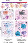
Adipose tissue microenvironments during aging: Effects on stimulated lipolysis
- PMID: 35131468
- PMCID: PMC8986088
- DOI: 10.1016/j.bbalip.2022.159118
Adipose tissue microenvironments during aging: Effects on stimulated lipolysis
Abstract
Adipose tissue is a critical organ for nutrient sensing, energy storage and maintaining metabolic health. The failure of adipose tissue homeostasis leads to metabolic disease that is seen during obesity or aging. Local metabolic processes are coordinated by interacting microenvironments that make up the complexity and heterogeneity of the adipose tissue. Catecholamine-induced lipolysis, a critical pathway in adipocytes that drives the release of stored triglyceride as free fatty acid after stimulation, is impaired during aging. The impairment of this pathway is associated with a failure to maintain a healthy body weight, core body-temperature during cold stress or mount an immune response. Along with impairments in aged adipocytes, aging is associated with an accumulation of inflammation, immune cell activation, and increased dysfunction in the nervous and lymphatic systems within the adipose tissue. Together these microenvironments support the initiation of stimulated lipolysis and the transport of free fatty acid under conditions of metabolic homeostasis. However, during aging, the defects in these cellular systems result in a reduction in ability to stimulate lipolysis. This review will focus on how the immune, nervous and lymphatic systems interact during tissue homeostasis, review areas that are impaired with aging and discuss areas of research that are currently unclear.
Keywords: Adipose tissue lipolysis; Aging; Immune cells; Inflammation; Lymphatic vessels; Sympathetic nervous system.
Copyright © 2022 Elsevier B.V. All rights reserved.
Conflict of interest statement
Declaration of interests
The authors declare that they have no known competing financial interests or personal relationships that could have appeared to influence the work reported in this paper.
Figures


Similar articles
-
Increased lipolysis in adipose tissues is associated with elevation of systemic free fatty acids and insulin resistance in perilipin null mice.Horm Metab Res. 2010 Apr;42(4):247-53. doi: 10.1055/s-0029-1243599. Epub 2010 Jan 20. Horm Metab Res. 2010. PMID: 20091459
-
Repetitive amiodarone administration causes liver damage via adipose tissue ER stress-dependent lipolysis, leading to hepatotoxic free fatty acid accumulation.Am J Physiol Gastrointest Liver Physiol. 2021 Sep 1;321(3):G298-G307. doi: 10.1152/ajpgi.00458.2020. Epub 2021 Jul 14. Am J Physiol Gastrointest Liver Physiol. 2021. PMID: 34259586
-
[Adipose triglyceride lipase regulates adipocyte lipolysis].Sheng Li Ke Xue Jin Zhan. 2008 Jan;39(1):10-4. Sheng Li Ke Xue Jin Zhan. 2008. PMID: 18357681 Review. Chinese.
-
Hormonal regulation of intracellular lipolysis in C57BL/6J mice: effect of diet-induced adiposity and data normalization.Metabolism. 2008 Oct;57(10):1405-13. doi: 10.1016/j.metabol.2008.05.010. Metabolism. 2008. PMID: 18803946
-
Adipose tissue lipolysis as a metabolic pathway to define pharmacological strategies against obesity and the metabolic syndrome.Pharmacol Res. 2006 Jun;53(6):482-91. doi: 10.1016/j.phrs.2006.03.009. Epub 2006 Mar 27. Pharmacol Res. 2006. PMID: 16644234 Review.
Cited by
-
Role for BLT1 in regulating inflammation within adipose tissue immune cells of aged mice.Immun Ageing. 2024 Aug 26;21(1):57. doi: 10.1186/s12979-024-00461-0. Immun Ageing. 2024. PMID: 39187841 Free PMC article.
-
Bariatric Surgery and Vitamin D: Trends in Older Women and Association with Clinical Features and VDR Gene Polymorphisms.Nutrients. 2023 Feb 4;15(4):799. doi: 10.3390/nu15040799. Nutrients. 2023. PMID: 36839157 Free PMC article.
-
Age-associated accumulation of B cells promotes macrophage inflammation and inhibits lipolysis in adipose tissue during sepsis.Cell Rep. 2024 Mar 26;43(3):113967. doi: 10.1016/j.celrep.2024.113967. Epub 2024 Mar 15. Cell Rep. 2024. PMID: 38492219 Free PMC article.
-
The Relaxin-3 Receptor, RXFP3, Is a Modulator of Aging-Related Disease.Int J Mol Sci. 2022 Apr 15;23(8):4387. doi: 10.3390/ijms23084387. Int J Mol Sci. 2022. PMID: 35457203 Free PMC article. Review.
-
B-cell interleukin 1 receptor 1 modulates the female adipose tissue immune microenvironment during aging.J Leukoc Biol. 2025 Feb 13;117(2):qiae219. doi: 10.1093/jleuko/qiae219. J Leukoc Biol. 2025. PMID: 39378334 Free PMC article.
References
-
- Scherer PE, Adipose tissue: from lipid storage compartment to endocrine organ. Diabetes 55, 1537–1545 (2006). - PubMed
Publication types
MeSH terms
Substances
Grants and funding
LinkOut - more resources
Full Text Sources

