

Laryngeal tuberculosis: a neglected diagnosis
- PMID: 35131802
- PMCID: PMC8823138
- DOI: 10.1136/bcr-2021-248095
Laryngeal tuberculosis: a neglected diagnosis
Abstract
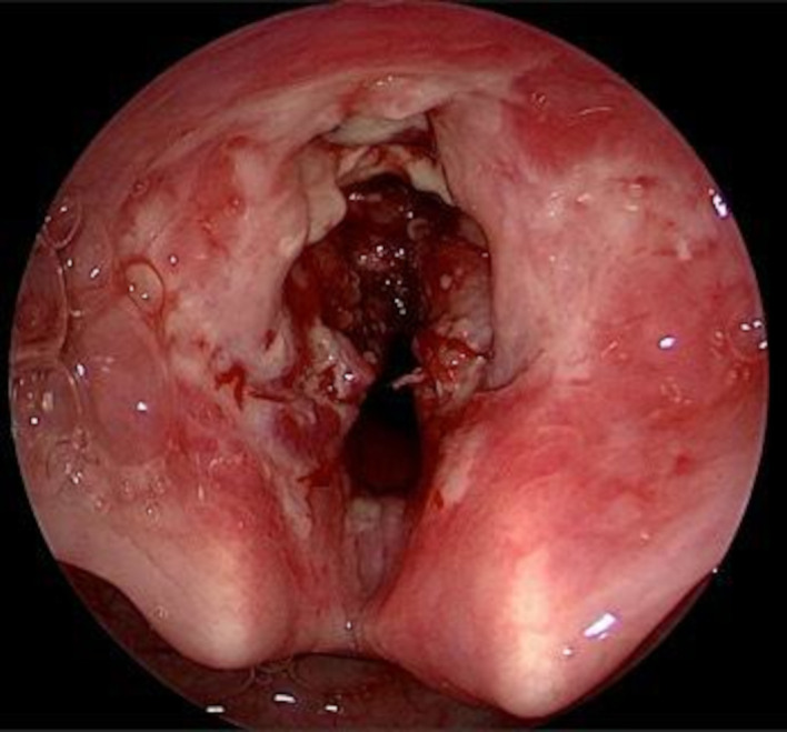
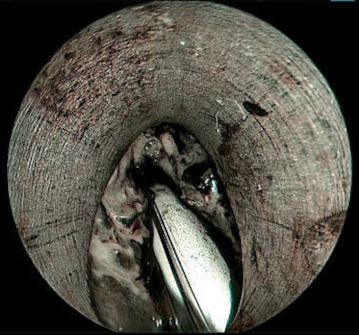
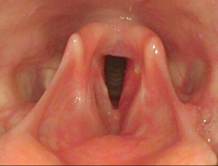
A 24-year-old woman visited the Ear Nose Throat (ENT) outpatient department with complaints of hoarseness for 2 months not responding to conservative management. Laryngoscopic examination revealed a whitish ulceroproliferative lesion in the anterior commissure and anterior two-thirds of bilateral true vocal cords with surrounding necrosis. In view of the above findings, the patient was planned for biopsy under general anaesthesia. Intraoperative findings showed multiple whitish necrotic friable tissue involving anterior two-thirds of bilateral false vocal cords, ventricle, bilateral true vocal cords, both aryepiglottic folds and laryngeal surface of epiglottis. Postoperative histopathology was consistent with tuberculosis. A pulmonology consultation was taken, and the patient was started on anti-tuberculosis chemotherapy. One month post therapy, the voice was symptomatically better. A flexible fibreoptic laryngoscopic examination was done, which revealed almost complete resolution of the lesion with minimal ulceration at the anterior one-third of right true vocal cord.
Keywords: TB and other respiratory infections; otolaryngology / ENT.
© BMJ Publishing Group Limited 2022. No commercial re-use. See rights and permissions. Published by BMJ.
Conflict of interest statement
Competing interests: None declared.
Figures


References
-
- Hirano M. Clinical examination of voice. New York, NY: Springer-Verlag, 1981.
Publication types
MeSH terms
LinkOut - more resources
Full Text Sources
Research Materials
Miscellaneous