

Universal stabilization of the influenza hemagglutinin by structure-based redesign of the pH switch regions
- PMID: 35131851
- PMCID: PMC8833195
- DOI: 10.1073/pnas.2115379119
Universal stabilization of the influenza hemagglutinin by structure-based redesign of the pH switch regions
Abstract
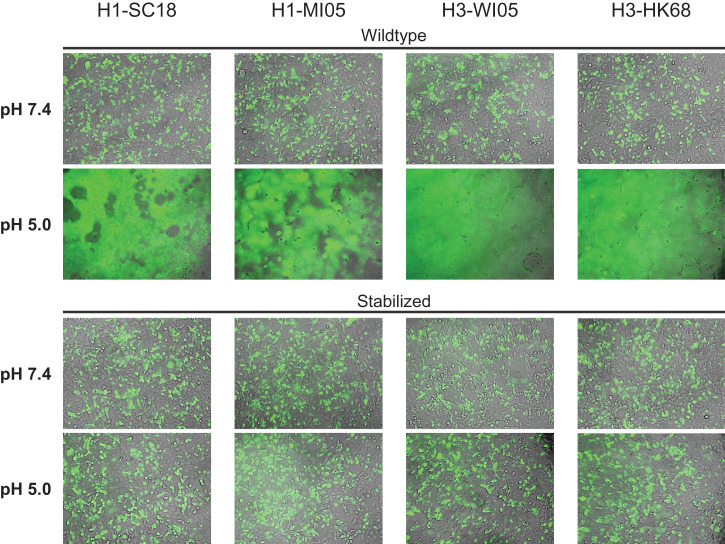
For an efficacious vaccine immunogen, influenza hemagglutinin (HA) needs to maintain a stable quaternary structure, which is contrary to the inherently dynamic and metastable nature of class I fusion proteins. In this study, we stabilized HA with three substitutions within its pH-sensitive regions where the refolding starts. An X-ray structure reveals how these substitutions stabilize the intersubunit β-sheet in the base and form an interprotomeric aliphatic layer across the stem while the native prefusion HA fold is retained. The identification of the stabilizing substitutions increases our understanding of how the pH sensitivity is structurally accomplished in HA and possibly other pH-sensitive class I fusion proteins. Our stabilization approach in combination with the occasional back mutation of rare amino acids to consensus results in well-expressing stable trimeric HAs. This repair and stabilization approach, which proves broadly applicable to all tested influenza A HAs of group 1 and 2, will improve the developability of influenza vaccines based on different types of platforms and formats and can potentially improve efficacy.
Keywords: fusion; influenza; protein design; protein stability; vaccine.
Copyright © 2022 the Author(s). Published by PNAS.
Conflict of interest statement
Competing interest statement: F.J.M., M.J., T.R., B.B., and J.P.M.L. are coinventors on related vaccine patents. F.J.M., M.J., T.R., P.B., I.J.M.B., M.d.M., D.V., L.L., B.K., M.J.G.B., J.J., B.B., and J.P.M.L. are employees of Janssen Vaccines & Prevention. M.J., J.J., B.B., P.B., and J.P.M.L. hold stock of Johnson & Johnson.
Figures
Similar articles
-
Hemagglutinin Stability Regulates H1N1 Influenza Virus Replication and Pathogenicity in Mice by Modulating Type I Interferon Responses in Dendritic Cells.J Virol. 2020 Jan 17;94(3):e01423-19. doi: 10.1128/JVI.01423-19. Print 2020 Jan 17. J Virol. 2020. PMID: 31694942 Free PMC article.
-
Influenza hemagglutinin (HA) stem region mutations that stabilize or destabilize the structure of multiple HA subtypes.J Virol. 2015 Apr;89(8):4504-16. doi: 10.1128/JVI.00057-15. Epub 2015 Feb 4. J Virol. 2015. PMID: 25653452 Free PMC article.
-
Production and stabilization of the trimeric influenza hemagglutinin stem domain for potentially broadly protective influenza vaccines.Proc Natl Acad Sci U S A. 2014 Jan 7;111(1):125-30. doi: 10.1073/pnas.1308701110. Epub 2013 Dec 16. Proc Natl Acad Sci U S A. 2014. PMID: 24344259 Free PMC article.
-
Acid-induced membrane fusion by the hemagglutinin protein and its role in influenza virus biology.Curr Top Microbiol Immunol. 2014;385:93-116. doi: 10.1007/82_2014_393. Curr Top Microbiol Immunol. 2014. PMID: 25007844 Free PMC article. Review.
-
Influenza virus hemagglutinin stalk-based antibodies and vaccines.Curr Opin Virol. 2013 Oct;3(5):521-30. doi: 10.1016/j.coviro.2013.07.007. Epub 2013 Aug 24. Curr Opin Virol. 2013. PMID: 23978327 Free PMC article. Review.
Cited by
-
Pre-existing H1N1 immunity reduces severe disease with bovine H5N1 influenza virus.bioRxiv [Preprint]. 2024 Oct 23:2024.10.23.619881. doi: 10.1101/2024.10.23.619881. bioRxiv. 2024. PMID: 39484442 Free PMC article. Preprint.
-
Ferritin-Based HA DNA Vaccine Outperforms Conventional Designs in Inducing Protective Immunity Against Seasonal Influenza.Vaccines (Basel). 2025 Jul 10;13(7):745. doi: 10.3390/vaccines13070745. Vaccines (Basel). 2025. PMID: 40733722 Free PMC article.
-
Immunogenic and Protective Properties of Recombinant Hemagglutinin of Influenza A (H5N8) Virus.Vaccines (Basel). 2024 Jan 29;12(2):143. doi: 10.3390/vaccines12020143. Vaccines (Basel). 2024. PMID: 38400127 Free PMC article.
-
Hemagglutinin glycosylation pattern-specific effects: implications for the fitness of H9.4.2.5-branched H9N2 avian influenza viruses.Emerg Microbes Infect. 2024 Dec;13(1):2364736. doi: 10.1080/22221751.2024.2364736. Epub 2024 Jun 14. Emerg Microbes Infect. 2024. PMID: 38847071 Free PMC article.
-
Development of an Influenza/COVID-19 Combination mRNA Vaccine Containing a Novel Multivalent Antigen Design That Enhances Immunogenicity of Influenza Virus B Hemagglutinins.Vaccines (Basel). 2025 Jun 11;13(6):628. doi: 10.3390/vaccines13060628. Vaccines (Basel). 2025. PMID: 40573959 Free PMC article.
References
Publication types
MeSH terms
Substances
LinkOut - more resources
Full Text Sources
Other Literature Sources
