

Small-Molecule MMRi62 Induces Ferroptosis and Inhibits Metastasis in Pancreatic Cancer via Degradation of Ferritin Heavy Chain and Mutant p53
- PMID: 35131878
- PMCID: PMC10258866
- DOI: 10.1158/1535-7163.MCT-21-0728
Small-Molecule MMRi62 Induces Ferroptosis and Inhibits Metastasis in Pancreatic Cancer via Degradation of Ferritin Heavy Chain and Mutant p53
Abstract
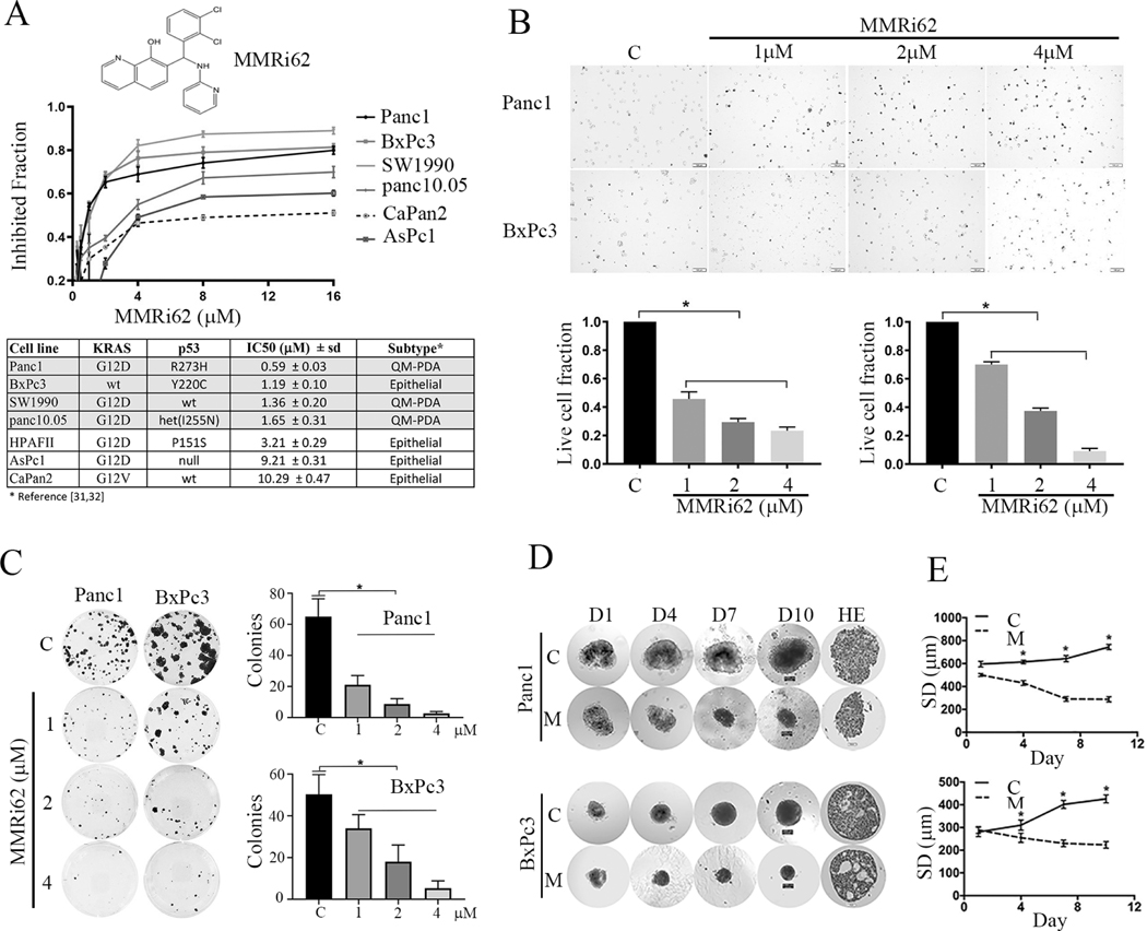
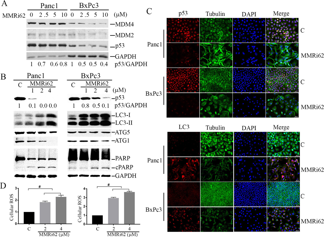
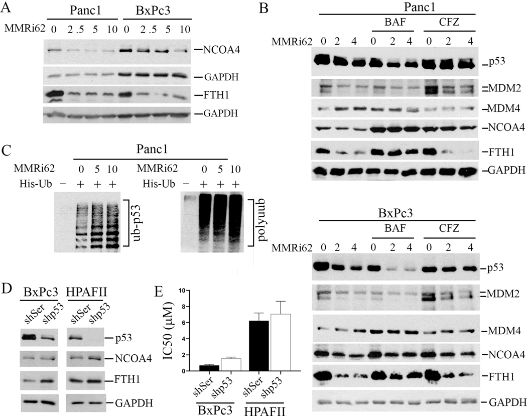
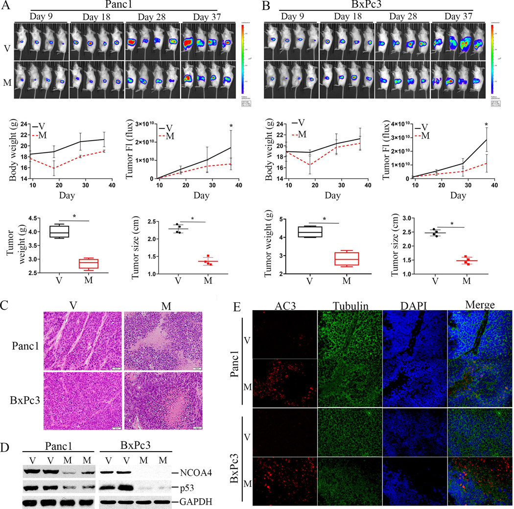
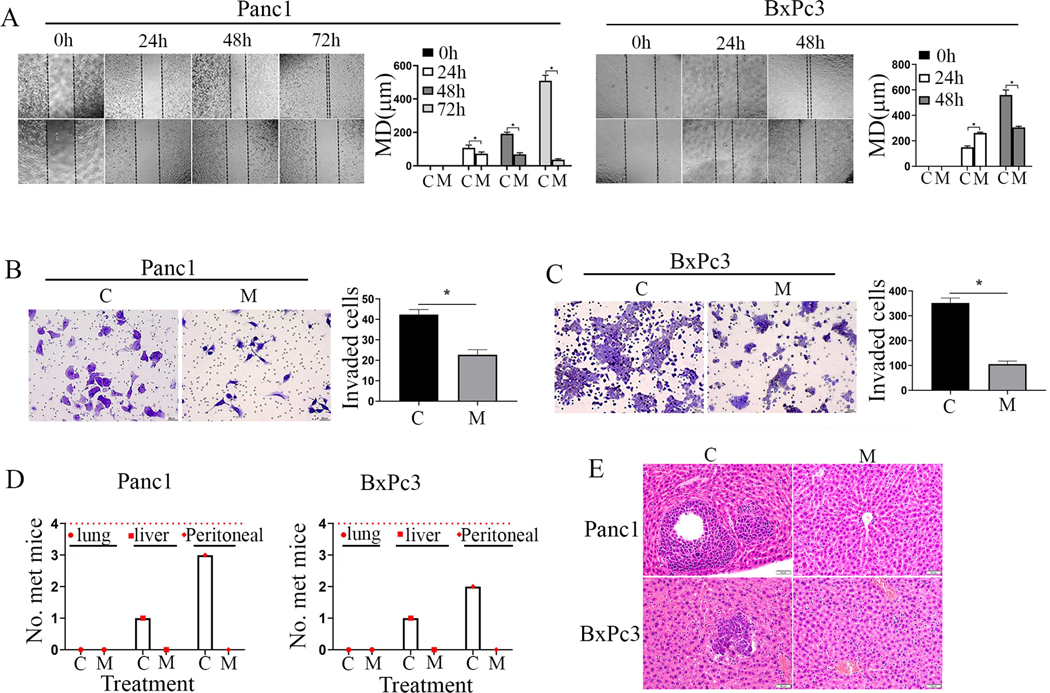
High frequency of KRAS and TP53 mutations is a unique genetic feature of pancreatic ductal adenocarcinoma (PDAC). TP53 mutation not only renders PDAC resistance to chemotherapies but also drives PDAC invasiveness. Therapies targeting activating mutant KRAS are not available and the outcomes of current PDAC treatment are extremely poor. Here, we report that MMRi62, initially identified as an MDM2-MDM4-targeting small molecule with p53-independent pro-apoptotic activity, shows anti-PDAC activity in vitro and in vivo. We show that MMRi62 inhibits proliferation, clonogenic, and spheroid growth of PDAC cells by induction of cell death. MMRi62-induced cell death in PDAC is characteristic of ferroptosis that is associated with increased autophagy, increased reactive oxygen species, and lysosomal degradation of NCOA4 and ferritin heavy chain (FTH1). In addition to induced degradation of FTH1, MMRi62 also induces proteasomal degradation of mutant p53. Interestingly, MMRi62-induced ferroptosis occurs in PDAC cell lines harboring either KRAS and TP53 double mutations or single TP53 mutation. In orthotopic xenograft PDAC mouse models, MMRi62 was capable of inhibiting tumor growth in mice associated with downregulation of NCOA4 and mutant p53 in vivo. Strikingly, MMRi62 completely abrogated metastasis of orthotopic tumors to distant organs, which is consistent with MMRi62's ability to inhibit cell migration and invasion in vitro. These findings identified MMRi62 as a novel ferroptosis inducer capable of suppressing PDAC growth and overcoming metastasis.
©2022 American Association for Cancer Research.
Conflict of interest statement
Disclosure of Potential Conflicts of Interest
No potential conflicts of interest are declared by authors.
Figures
Similar articles
-
MGST1 facilitates novel KRASG12D inhibitor resistance in KRASG12D-mutated pancreatic ductal adenocarcinoma by inhibiting ferroptosis.Mol Med. 2024 Nov 5;30(1):199. doi: 10.1186/s10020-024-00972-y. Mol Med. 2024. PMID: 39501138 Free PMC article.
-
Identification of TPI1 As a potential therapeutic target in pancreatic cancer with dependency of TP53 mutation using multi-omics analysis.Cancer Sci. 2024 Nov;115(11):3622-3635. doi: 10.1111/cas.16302. Epub 2024 Sep 11. Cancer Sci. 2024. PMID: 39259678 Free PMC article.
-
Docosahexaenoic acid inhibits the proliferation of Kras/TP53 double mutant pancreatic ductal adenocarcinoma cells through modulation of glutathione level and suppression of nucleotide synthesis.PLoS One. 2020 Nov 2;15(11):e0241186. doi: 10.1371/journal.pone.0241186. eCollection 2020. PLoS One. 2020. PMID: 33137095 Free PMC article.
-
Emerging Potential Mechanism and Therapeutic Target of Ferroptosis in PDAC: A Promising Future.Int J Mol Sci. 2022 Nov 30;23(23):15031. doi: 10.3390/ijms232315031. Int J Mol Sci. 2022. PMID: 36499358 Free PMC article. Review.
-
Cell death in pancreatic cancer: from pathogenesis to therapy.Nat Rev Gastroenterol Hepatol. 2021 Nov;18(11):804-823. doi: 10.1038/s41575-021-00486-6. Epub 2021 Jul 30. Nat Rev Gastroenterol Hepatol. 2021. PMID: 34331036 Review.
Cited by
-
Effects of TP53 Mutations and miRs on Immune Responses in the Tumor Microenvironment Important in Pancreatic Cancer Progression.Cells. 2022 Jul 9;11(14):2155. doi: 10.3390/cells11142155. Cells. 2022. PMID: 35883598 Free PMC article. Review.
-
From mechanisms to medicine: Ferroptosis as a Therapeutic target in liver disorders.Cell Commun Signal. 2025 Mar 7;23(1):125. doi: 10.1186/s12964-025-02121-2. Cell Commun Signal. 2025. PMID: 40055721 Free PMC article. Review.
-
Dual Targeting of MDM4 and FTH1 by MMRi71 for Induced Protein Degradation and p53-Independent Apoptosis in Leukemia Cells.Molecules. 2022 Nov 8;27(22):7665. doi: 10.3390/molecules27227665. Molecules. 2022. PMID: 36431769 Free PMC article.
-
Small molecule MMRi62 targets MDM4 for degradation and induces leukemic cell apoptosis regardless of p53 status.Front Oncol. 2022 Aug 5;12:933446. doi: 10.3389/fonc.2022.933446. eCollection 2022. Front Oncol. 2022. PMID: 35992795 Free PMC article.
-
Ursolic Acid induces ferroptosis by affecting redox balance and FADS2-mediated unsaturated fatty acid synthesis in Non-Small Cell Lung Cancer.J Cancer. 2025 May 27;16(8):2553-2566. doi: 10.7150/jca.105863. eCollection 2025. J Cancer. 2025. PMID: 40535804 Free PMC article.
References
-
- Siegel RL, Miller KD, Fuchs HE, Jemal A. Cancer Statistics, 2021. CA Cancer J Clin. 2021;71:7–33. - PubMed
-
- Chen W, Zheng R, Baade PD, Zhang S, Zeng H, Bray F, et al. Cancer statistics in China, 2015. CA Cancer J Clin. 2016;66:115–32. - PubMed
-
- Rahib L, Smith BD, Aizenberg R, Rosenzweig AB, Fleshman JM, Matrisian LM. Projecting cancer incidence and deaths to 2030: the unexpected burden of thyroid, liver, and pancreas cancers in the United States. Cancer Res. 2014;74:2913–21. - PubMed
-
- Kleeff J, Korc M, Apte M, La Vecchia C, Johnson CD, Biankin AV, et al. Pancreatic cancer. Nat Rev Dis Primers. 2016;2:16022. - PubMed
-
- Neoptolemos JP, Kleeff J, Michl P, Costello E, Greenhalf W, Palmer DH. Therapeutic developments in pancreatic cancer: current and future perspectives. Nat Rev Gastroenterol Hepatol. 2018;15:333–48. - PubMed
Publication types
MeSH terms
Substances
Grants and funding
LinkOut - more resources
Full Text Sources
Other Literature Sources
Medical
Research Materials
Miscellaneous
