. 2022 Feb 15;119(7):e2112696119.
doi: 10.1073/pnas.2112696119.

Intravesical delivery of KDM6A-mRNA via mucoadhesive nanoparticles inhibits the metastasis of bladder cancer

Affiliations

Intravesical delivery of KDM6A-mRNA via mucoadhesive nanoparticles inhibits the metastasis of bladder cancer

Na Kong et al. Proc Natl Acad Sci U S A. .

Abstract

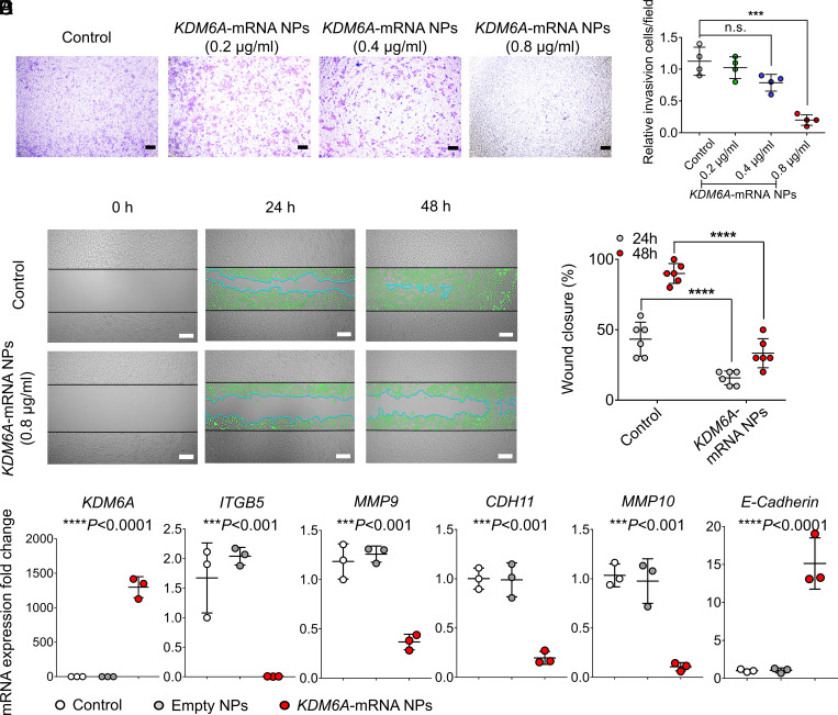
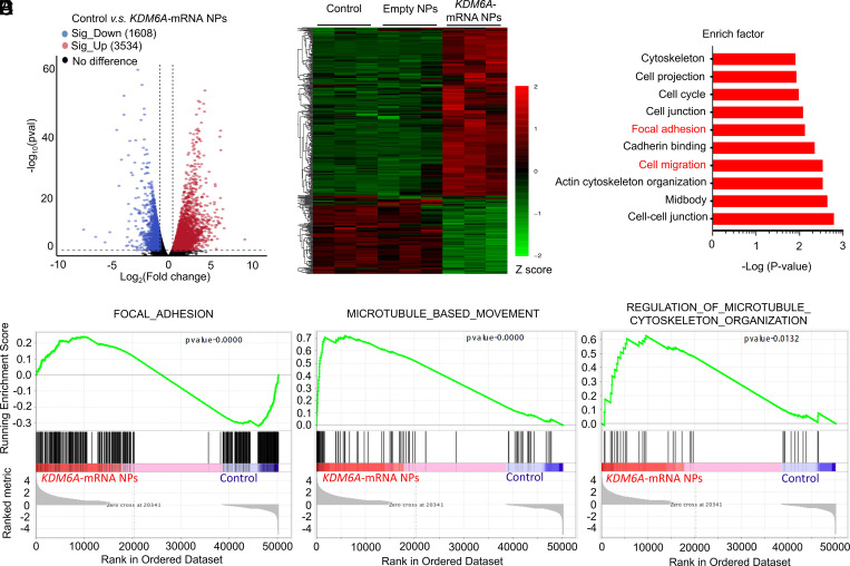
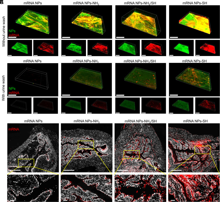
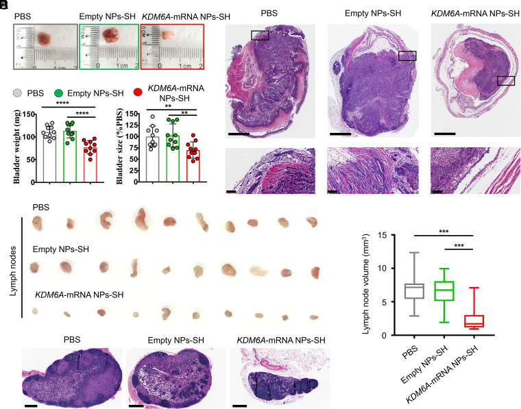
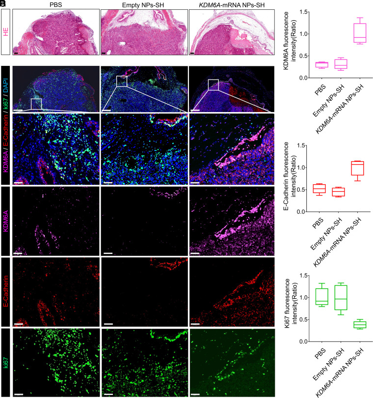
Lysine-specific demethylase 6A (KDM6A), also named UTX, is frequently mutated in bladder cancer (BCa). Although known as a tumor suppressor, KDM6A's therapeutic potential in the metastasis of BCa remains elusive. It also remains difficult to fulfill the effective up-regulation of KDM6A levels in bladder tumor tissues in situ to verify its potential in treating BCa metastasis. Here, we report a mucoadhesive messenger RNA (mRNA) nanoparticle (NP) strategy for the intravesical delivery of KDM6A-mRNA in mice bearing orthotopic Kdm6a-null BCa and show evidence of KDM6A's therapeutic potential in inhibiting the metastasis of BCa. Through this mucoadhesive mRNA NP strategy, the exposure of KDM6A-mRNA to the in situ BCa tumors can be greatly prolonged for effective expression, and the penetration can be also enhanced by adhering to the bladder for sustained delivery. This mRNA NP strategy is also demonstrated to be effective for combination cancer therapy with other clinically approved drugs (e.g., elemene), which could further enhance therapeutic outcomes. Our findings not only report intravesical delivery of mRNA via a mucoadhesive mRNA NP strategy but also provide the proof-of-concept for the usefulness of these mRNA NPs as tools in both mechanistic understanding and translational study of bladder-related diseases.

Keywords: KDM6A; bladder cancer; elemene; intravesical delivery; mRNA nanoparticles.


Conflict of interest statement

The authors declare no competing interest.

Figures

Fig. 1.
Fig. 1.
On-demand introduction of exogenous and functional proteins in Kdm6a-null BCa cells via mRNA NPs. (A) mRNA NP morphology was observed through TEM (Scale bar, 200 nm). (B) Stability of the mRNA NPs in PBS containing 10% serum throughout 72 h. The NP diameters were confirmed through dynamic light scattering measurements. (C) CLSM imaging of Kdm6a-null KU19-19 cells after different duration of incubation (1, 3, and 6 h) with Cy5-mRNA NPs (red). CellLight Late Endosomes-GFP was used to stain endosomes (green), and Hoechst 33342 was utilized to stain nuclei (blue). Cells after 6 h of incubation with free Cy5-mRNA were assigned to the control group (Scale bars, 10 µm). (D) IF staining of KDM6A in Kdm6a-null KU19-19 cells after treatment with empty NPs or KDM6A-mRNA NPs (Scale bars, 50 µm).
Fig. 2.
Fig. 2.
mRNA NPs restores KDM6A in Kdm6a-null BCa cells, inhibits metastasis-related pathways, and prevents the invasion and migration of BCa cells in vitro. (A) Representative images of Kdm6a-null KU19-19 BCa cells after treatment with KDM6A-mRNA NPs at different concentrations in the 24-transwell system from transwell invasion assays. KU19-19 cells without NP treatment were assigned to the control group (Scale bars, 160 µm). (B) Quantitative analysis of the transwell invasion assays. Invasive cells were counted in five representative magnified views per transwell. Experiments were carried out four times. Statistical significance was defined by ***P < 0.001. (C) Representative images of Kdm6a-null KU19-19 cells with or without treatment with KDM6A-mRNA NPs from in vitro wound healing assays (incubation time: 24 and 48 h). Green cells represent invasive and migrating cells. Blue lines represent the borderlines (Scale bars, 200 µm). (D) Quantitative analysis of the in vitro wound healing assays. Statistical significance was defined by ****P < 0.0001. (E) qRT-PCR was carried out to measure the expression of KDM6A, ITGB5, MMP9, CDH11, MMP10, and E-Cadherin. Statistical significance was defined as ***P < 0.001 and ****P < 0.0001 between control group and KDM6A-mRNA NPs Group.
Fig. 3.
Fig. 3.
Transcriptomic analysis between Kdm6a-null KU19-19 cells (control group) and KDM6A-mRNA NP–treated Kdm6a-null KU19-19 cells (KDM6A-mRNA NPs Group). (A) Volcano plot of gene expression (Control versus KDM6A-mRNA NPs; fold changes ≥ 2; P < 0.05). (B) Hierarchical clustering and heatmap of representative gene expression resulting from control group, Empty NPs Group (i.e., empty NP-treated Kdm6a-null KU19-19 cells), and KDM6A-mRNA NPs Group through transcriptomic sequencing. Row-scaled z-scores of quantile-normalized gene expression (from >5,000 genes; P < 0.0001; Green: down-regulated genes; Red: up-regulated genes). (C) KEGG pathway analysis on the significant gene expression. (D) GSEA of the genes associated with the focal adhesion signaling (P < 0.0001) in control group and KDM6A-mRNA NPs Group. (E) GSEA of the genes associated with the microtubule-based movement signaling (P < 0.0001) in control group and KDM6A-mRNA NPs Group. (F) GSEA of the genes associated with the regulation of microtubule cytoskeleton organization signaling (P = 0.0132) in control group and KDM6A-mRNA NPs Group.
Fig. 4.
Fig. 4.
Engineering of surface mucoadhesive mRNA NPs for effective intravesical delivery of mRNA. (A) CLSM volume view images of mouse bladder walls after incubation (2 h) with nonmucoadhesive mRNA NPs, mucoadhesive mRNA NPs-NH2, mucoadhesive mRNA NPs-NH2/SH, or mucoadhesive mRNA NPs-SH. mRNA was labeled with Cy5 (red fluorescence), and NPs were labeled with Fluorescein (FITC) (green fluorescence) (Scale bar, 400 μm). (B) CLSM volume view images of mouse bladder walls after 2 h of incubation with different mRNA NPs, followed by another 3 h of incubation in urine (i.e., urine wash). mRNA was labeled with Cy5 (red fluorescence), and NPs were labeled with FITC (green fluorescence) (Scale bar, 400 μm). (C) Sections of the bladder tissues from mice treating with nonmucoadhesive mRNA NPs, mucoadhesive mRNA NPs-NH2, mucoadhesive mRNA NPs-NH2/SH, or mucoadhesive mRNA NPs-SH via intravesical administration (Scale bars in raw images, 500 µm; Scale bars in enlarged images, 50 µm). mRNA was labeled with Cy5 (red fluorescence).
Fig. 5.
Fig. 5.
Introduction of KDM6A via mucoadhesive mRNA NPs in mice bearing orthotopic BCa tumors could inhibit metastasis potential of BCa. (A) Representative images of the entire bladders bearing orthotopic primary BCa tumors in different groups. (B) The weight (mg) and the size changes (% compared with the PBS group) of the harvested bladders from different groups. Data shown as means ± SEM (n = 10) with a two-tailed Student’s t test (**P < 0.01, ****P < 0.0001). (C) Representative H&E staining sections from the entire bladders bearing orthotopic primary BCa tumors in different groups (Scale bars in raw images, 1.25 mm; Scale bars in enlarged images, 100 µm). (D) Representative images of the maximum-volume lymph nodes collected from each group (n = 10). (E) Histogram analysis of the volume calculated from all the lymph nodes in different groups. Data shown as means ± SEM (n = 10) with a two-tailed Student’s t test (***P < 0.001). (F) Representative H&E staining images of the lymph nodes from different groups (Scale bars, 400 μm).
Fig. 6.
Fig. 6.
Local restoration of KDM6A in Kdm6a-null BCa tumors via a mucoadhesive mRNA NP–mediated strategy for the mechanistic understanding of KDM6A’s role in inhibiting BCa metastasis in vivo. (A) H&E staining sections of the bladders bearing orthotopic primary BCa tumors that used for the following IF analysis (Scale bars, 200 µm). (B) IF analysis of the KDM6A (pink), E-Cadherin (red), and Ki67 (green) expression in orthotopic Kdm6a-null KU19-19 tumors after different treatments (Scale bars in raw images, 200 µm; Scale bars in enlarged images, 50 µm). The nucleus was stained by DAPI (blue). Quantitative analysis of (C) KDM6A (pink), (D) E-Cadherin (red), and (E) Ki67 (green) expression in the bladders bearing orthotopic primary BCa tumors from different groups.

Comment in

  • Urolithiasis/Endourology.
    Assimos DG. Assimos DG. J Urol. 2022 Nov;208(5):1154-1155. doi: 10.1097/JU.0000000000002913. Epub 2022 Aug 17. J Urol. 2022. PMID: 35975568 No abstract available.
  • Uro-Science.
    Atala A. Atala A. J Urol. 2022 Nov;208(5):1152-1153. doi: 10.1097/JU.0000000000002932. Epub 2022 Aug 22. J Urol. 2022. PMID: 35993121 No abstract available.

References

    1. Roy D. M., Walsh L. A., Chan T. A., Driver mutations of cancer epigenomes. Protein Cell 5, 265–296 (2014). - PMC - PubMed
    1. Dunford A., et al. , Tumor-suppressor genes that escape from X-inactivation contribute to cancer sex bias. Nat. Genet. 49, 10–16 (2017). - PMC - PubMed
    1. Ler L. D., et al. , Loss of tumor suppressor KDM6A amplifies PRC2-regulated transcriptional repression in bladder cancer and can be targeted through inhibition of EZH2. Sci. Transl. Med. 9, eaai8312 (2017). - PubMed
    1. Kaneko S., Li X., X chromosome protects against bladder cancer in females via a KDM6A-dependent epigenetic mechanism. Sci. Adv. 4, eaar5598 (2018). - PMC - PubMed
    1. van Haaften G., et al. , Somatic mutations of the histone H3K27 demethylase gene UTX in human cancer. Nat. Genet. 41, 521–523 (2009). - PMC - PubMed

MeSH terms

LinkOut - more resources