

Does computed tomography-guided percutaneous catheter drainage is effective for spinal tuberculous abscess: a midterm results
- PMID: 35132064
- PMCID: PMC8821638
- DOI: 10.1038/s41394-022-00488-9
Does computed tomography-guided percutaneous catheter drainage is effective for spinal tuberculous abscess: a midterm results
Abstract
Study design: Retrospective cohort study.
Purpose: To evaluate an effectiveness and report a midterm clinical outcome in pain and neurological status in spinal tuberculous abscess after treated by CT-guided percutaneous catheter drainage.
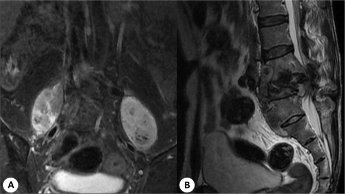
Overview of literature: Spinal tuberculosis is one of the destructive forms of tuberculosis infection, which can cause undesirable consequences. The gold standard of surgical treatment of spinal tuberculosis with tuberculous abscess is radical debridement, abscess drainage, and bone grafting of the defect via anterior approach. However, this treatment may lead to several serious complications. CT-guided percutaneous catheter drainage is an alternative procedure for this condition and may reduce the serious complications from standard surgical treatment.
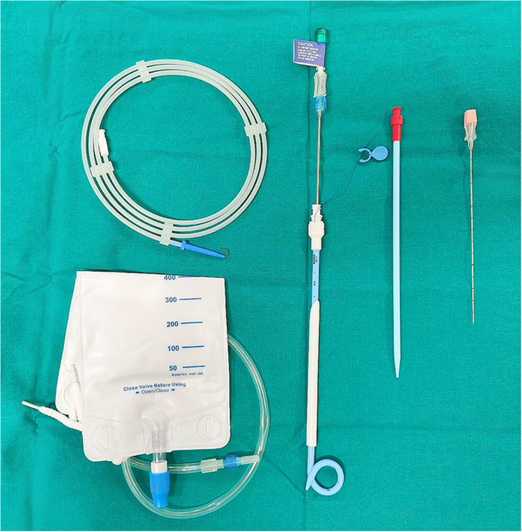
Materials and methods: The medical record of the patients with spinal tuberculosis with tuberculous abscess who underwent CT-guided percutaneous catheter drainage (CT-guided PCD) from 2015 to 2021. The visual analog pain scale (VAS), Frankel grading scale, duration of drainage, amount of spinal tuberculous abscess, and complications were evaluated.
Results: Twenty-nine patients (mean age 44 years old) were included in the study. All patients were followed up for 24 to 72 months with an average of 36 months. Level involvements were mostly found in L1-L2 followed by L2-L3 and T12-L1 levels. A 14-Fr catheter was the mostly use followed by 16-Fr catheter. Amount of abscess drainage ranged from 110 to 2,490 ml (mean 599 ml). The drainage duration ranged from 6 to 42 days (mean 17 days). Additional surgery was performed in three patients due to subsequent mechanical instability developed despite successful drainage of abscess. At the last follow-up, VAS, Frankel grading scale were improved significantly in all patients without complications.
Conclusions: CT-guided percutaneous catheter drainage is a safe and effective alternative procedure in the treatment of spinal tuberculous abscess patients with high success rate, less complications, and satisfied midterm outcomes.
© 2022. The Author(s), under exclusive licence to International Spinal Cord Society.
Conflict of interest statement
The authors declared no potential conflicts of interest with respect to the research, authorship, and/or publication of this article.
Figures


References
-
- World Health Organization. Global tuberculosis report 2020. WHO 2020; https://www.who.int/tb/publications/global_report/en/. Accessed 20 Jul 2021.
-
- Trecarichi EM, Meco ED, Mazzotta V, Fantoni M. Tuberculous spondylodiscitis: epidemiology, clinical features, treatment, and outcome. Eur Rev Med Pharm Sci. 2012;16(Suppl 2):S58–S72. - PubMed
Publication types
MeSH terms
LinkOut - more resources
Full Text Sources
Medical
