. 2022 Jan 29:2022:4566851.
doi: 10.1155/2022/4566851. eCollection 2022.

The ROS/GRK2/HIF-1 α/NLRP3 Pathway Mediates Pyroptosis of Fibroblast-Like Synoviocytes and the Regulation of Monomer Derivatives of Paeoniflorin

Affiliations

The ROS/GRK2/HIF-1 α/NLRP3 Pathway Mediates Pyroptosis of Fibroblast-Like Synoviocytes and the Regulation of Monomer Derivatives of Paeoniflorin

Zhongyang Hong et al. Oxid Med Cell Longev. .

Abstract

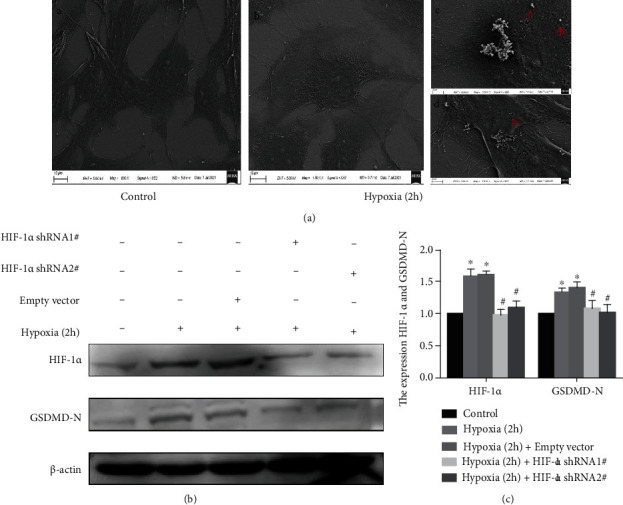
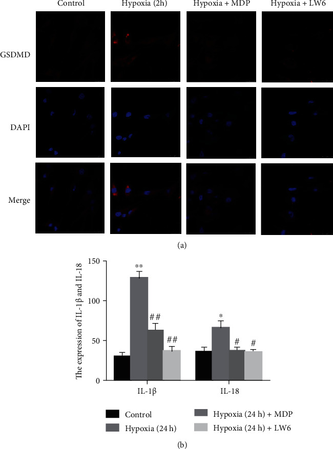
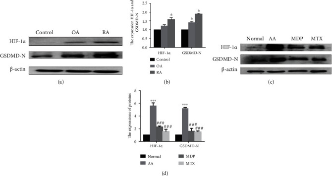
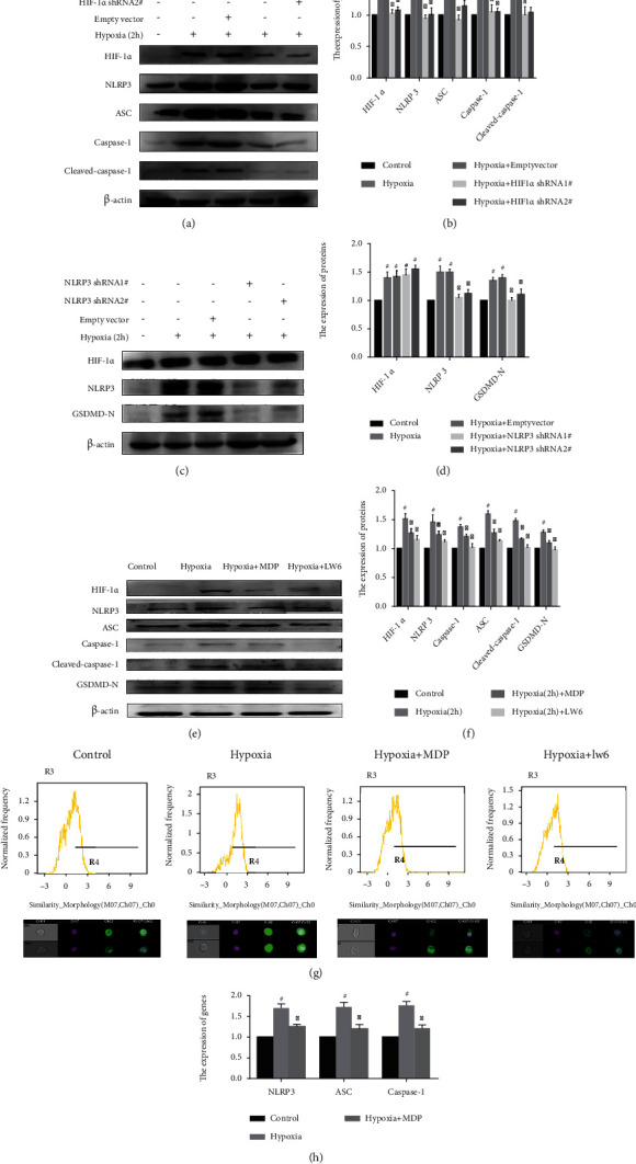
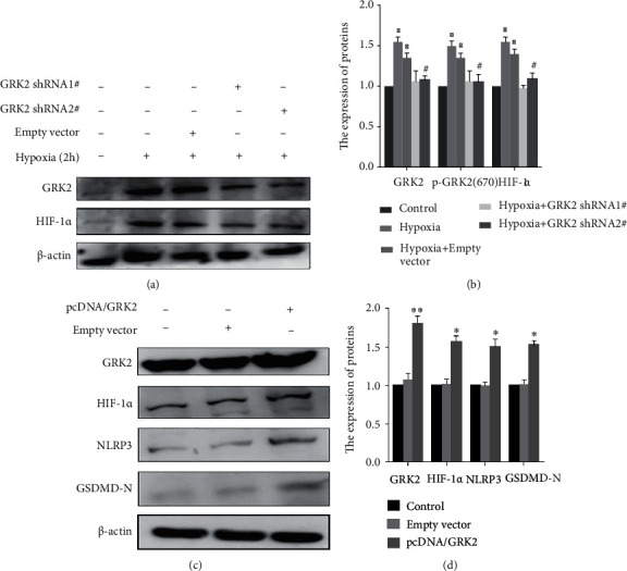
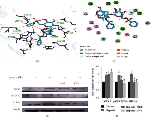
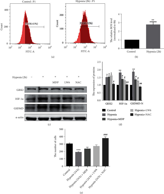
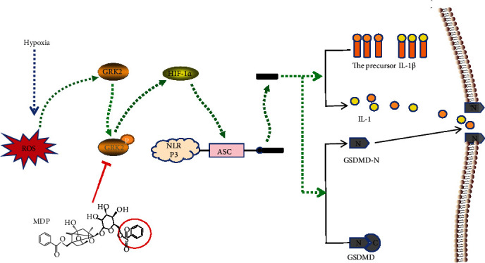
Hypoxia is an important factor in the development of synovitis in rheumatoid arthritis (RA). The previous study of the research group found that monomeric derivatives of paeoniflorin (MDP) can alleviate joint inflammation in adjuvant-induced arthritis (AA) rats by inhibiting macrophage pyroptosis. This study revealed increased levels of hypoxia-inducible factor- (HIF-) 1α and N-terminal p30 fragment of GSDMD (GSDMD-N) in fibroblast-like synoviocytes (FLS) of RA patients and AA rats, while MDP significantly inhibited their expression. Subsequently, FLS were exposed to a hypoxic environment or treated with cobalt ion in vitro. Western blot and immunofluorescence analysis showed increased expression of G protein-coupled receptor kinase 2 (GRK2), HIF-1α, nucleotide-binding oligomerization segment-like receptor family 3 (NLRP3), ASC, caspase-1, cleaved-caspase-1, and GSDMD-N. Electron microscopy revealed FLS pyroptosis after exposure in hypoxia. Next, corresponding shRNAs were transferred into FLS to knock down hypoxia-inducible factor- (HIF-) 1α, and in turn, NLRP3 and western blot results confirmed the same. The enhanced level of GSDMD was reversed under hypoxia by inhibiting NLRP3 expression. Knockdown and overexpression of GRK2 in FLS revealed GRK2 to be a positive regulator of HIF-1α. Levels of GRK2 and HIF-1α were inhibited by eliminating excess reactive oxygen species (ROS). Furthermore, MDP reduced FLS pyroptosis through targeted inhibition of GRK2 phosphorylation. According to these findings, hypoxia induces FLS pyroptosis through the ROS/GRK2/HIF-1α/NLRP3 pathway, while MDP regulates this pathway to reduce FLS pyroptosis.


Conflict of interest statement

The authors have declared no conflicts of interest.

Figures

Figure 1
Figure 1
Hypoxia-mediated induction of pyroptosis in fibroblast-like synoviocytes (FLS). (a) Representative scanning electron microscope images of FLS pyroptosis. a, b FLS cultured under normoxia and hypoxia, respectively (1000x). c, d FLS cultured under hypoxia for 2 h (3000x). A HIF-1α short hairpin (shHIF-1α) construct was transfected into FLS, and hypoxic condition was imposed for 2 h. (b, c) Immunoblot analysis and quantification of hypoxia-inducible factor (HIF)-1α and N-terminal domain of human gasdermin D (GSDMD-N). Data are presented as mean ± standard deviation. n = 3. p < 0.05 compared with the control group and #p < 0.05 compared with the empty vector group.
Figure 2
Figure 2
Monomeric derivatives of paeoniflorin (MDP) reduces hypoxia-induced fibroblast-like synoviocytes (FLS) pyroptosis. (a) Representative images of immunofluorescence of N-terminal domain of human gasdermin D (GSDMD-N) in FLS. (b) Enzyme-linked immunosorbent assay and quantification of interleukin- (IL-) 1β and IL-18 (n = 5). Data are expressed as mean + standard deviation. p < 0.05, ∗∗p < 0.01 vs. the control group; #p < 0.05, ##p < 0.01 vs. the hypoxia group.
Figure 3
Figure 3
The levels of hypoxia-inducible factor (HIF)-1α and N-terminal domain of human gasdermin D (GSDMD-N) increase in fibroblast-like synoviocytes (FLS) of RA patients and adjuvant-induced arthritis (AA) rats. (a, b) Immunoblot analysis and quantification of HIF-1α and GSDMD-N in the FLS of RA patients. (c, d) Immunoblot analysis of HIF-1α and GSDMD-N expression levels in FLS of normal, AA, AA+MDP, and AA+MTX rats. Data are presented as mean ± standard deviation. n = 3. p < 0.05, ∗∗∗p < 0.001 compared with the control group and ###p < 0.001 compared with the AA group (MTX: methotrexate; MDP: monomeric derivatives of paeoniflorin).
Figure 4
Figure 4
Hypoxia induces fibroblast-like synoviocytes (FLS) pyroptosis by upregulating the expression of nucleotide-binding oligomerization segment-like receptor family 3 (NLRP3) inflammasome in FLS and the effect of monomeric derivatives of paeoniflorin (MDP). (a, b) shHIF-1α was transfected into FLS and exposed to hypoxia for 2 h. The expression of hypoxia-inducible factor (HIF)-1α, NLRP3, speck-like protein containing CARD (ASC), caspase-1, and cleaved-caspase-1 in FLS was detected by Western blot (n = 3). (c, d) NLRP3 short hairpin (shNLRP3) construct was transfected into FLS and exposed to hypoxia for 2 h. The expression of HIF-1α, NLRP3, and N-terminal domain of human gasdermin D (GSDMD-N) in FLS after exposure to hypoxia for 2 h was detected by Western blot (n = 3). (e, f) FLS were treated with or without MDP and exposed to hypoxia for 2 h. The expression of HIF-1α, NLRP3, ASC, caspase-1, cleaved-caspase-1, and GSDMD-N in FLS was detected by Western blot (n = 3). Data are expressed as mean ± SD. #p < 0.05, ##p < 0.01 vs. the control group; p < 0.05, ∗∗p < 0.01 vs. the hypoxia group. (g) Analysis of expression levels of HIF-1α in the nucleus. (h) Quantitative PCR analysis and quantification of the mRNA levels of NLRP3, ASC, and caspase-1. Data are expressed as mean SD. #p < 0.05 vs. the control group; p < 0.05 vs. the hypoxia group.
Figure 5
Figure 5
G protein-coupled receptor kinase 2 (GRK2) promotes the expression of hypoxia-inducible factor (HIF)-1α. (a, b) shGRK2 was transfected into fibroblast-like synoviocytes (FLS) and exposed to hypoxia for 2 h. Immunoblot analysis and quantification of GRK2 and HIF-1α. (c, d) GRK2 overexpression plasmid was transfected into FLS. Immunoblot analysis and quantification of GRK2, HIF-1α, nucleotide-binding oligomerization segment-like receptor family 3 (NLRP3), and N-terminal domain of human gasdermin D (GSDMD-N). The values are shown as the mean ± standard deviation (n = 3). p < 0.05, ∗∗p < 0.01 compared with the control group and #p < 0.05 compared with the hypoxia group or the NC group.
Figure 6
Figure 6
Monomeric derivative of paeoniflorin (MDP) inhibits the expression of hypoxia-inducible factor (HIF)-1α through targeted inhibition of GRK2 S670 phosphorylation. (a) Molecular docking modeling of the compound MDP and G protein-coupled receptor kinase 2 (GRK2), the small molecule, and the critical interaction of 3KRW (human GRK2 in complex with Gbetgamma subunits and balanol) are represented by sticks. (b) A schematic representation of the binding mode of MDP in the GRK2 binding site of 3KRW. (c, d) FLS were treated with or without MDP and exposed to hypoxia for 2 h. Immunoblot analysis and quantification of GRK2, p-GRK2 (S670), and HIF-1α. The values are shown as the mean ± standard error of the mean (n = 3). p < 0.05 compared with the control group and #p < 0.05 compared with the hypoxia group.
Figure 7
Figure 7
Excessive reactive oxygen species (ROS) upregulates the expression level of G protein-coupled receptor kinase 2 (GRK2) under hypoxia in fibroblast-like synoviocytes (FLS). (a, b) The analysis and quantification of ROS in FLS. (c, d) FLS were pretreated with N-acetyl-l-cysteine (NAC) before exposure to hypoxia. Immunoblot analysis and quantification of GRK2, hypoxia-inducible factor (HIF)-1α, and N-terminal domain of human gasdermin D (GSDMD-N) in FLS. (e) Cell proliferation analysis.
Figure 8
Figure 8
Reactive oxygen species/G protein-coupled receptor kinase 2/hypoxia-inducible factor-1α/nucleotide-binding oligomerization segment-like receptor family 3- (ROS/GRK2/HIF-1α/NLRP3-) mediated pyroptosis in fibroblast-like synoviocytes (FLS) promotes synovitis under hypoxia and the regulation by monomeric derivatives of paeoniflorin (MDP). The level of ROS in FLS rises sharply under hypoxia. Then, excessive ROS promotes GRK2 expression and increases the levels of phosphorylated GRK2 S670, which then increases HIF-1α synthesis. This elevated HIF-1α is transferred to the nucleus to initiate the transcription of the NLRP3 inflammasome. The abnormally high level of NLRP3 inflammasome is activated under hypoxia to form activated cleaved-caspase-1, which, in turn, shears gasdermin D (GSDMD), interleukin- (IL-) 1β, and IL-18 to induce FLS pyroptosis. Monomeric derivative of paeoniflorin (MDP) inhibits the phosphorylation of GRK2 S670 to reduce FLS pyroptosis and relieve synovitis.

Similar articles

Cited by

References

    1. Yang M., Liu Y., Mo B., et al. Helios but not CD226, TIGIT and Foxp3 is a potential marker for CD4+ Treg cells in patients with rheumatoid arthritis. Cellular Physiology and Biochemistry . 2019;52(5):1178–1192. doi: 10.33594/000000080. - DOI - PMC - PubMed
    1. Moller B., Kollert F., Sculean A., Villiger P. M. Infectious triggers in periodontitis and the gut in rheumatoid arthritis (RA): a complex story about association and causality. Frontiers in Immunology . 2020;11:p. 1108. - PMC - PubMed
    1. Yu S., Lu Y., Zong M., Tan Q., Fan L. Hypoxia-induced miR-191-C/EBPβ signaling regulates cell proliferation and apoptosis of fibroblast-like synoviocytes from patients with rheumatoid arthritis. Arthritis Research & Therapy . 2019;21(1):p. 78. doi: 10.1186/s13075-019-1861-7. - DOI - PMC - PubMed
    1. Hu F., Mu R., Zhu J., et al. Hypoxia and hypoxia-inducible factor-1α provoke toll-like receptor signalling-induced inflammation in rheumatoid arthritis. Annals of the Rheumatic Diseases . 2014;73(5):928–936. doi: 10.1136/annrheumdis-2012-202444. - DOI - PubMed
    1. Kim J., Kim H. Y., Song S. Y., et al. Synergistic oxygen generation and reactive oxygen species scavenging by manganese ferrite/ceria co-decorated nanoparticles for rheumatoid arthritis treatment. ACS Nano . 2019;13(3):3206–3217. doi: 10.1021/acsnano.8b08785. - DOI - PubMed

MeSH terms