

Peroxiredoxin-5 Knockdown Accelerates Pressure Overload-Induced Cardiac Hypertrophy in Mice
- PMID: 35132351
- PMCID: PMC8817848
- DOI: 10.1155/2022/5067544
Peroxiredoxin-5 Knockdown Accelerates Pressure Overload-Induced Cardiac Hypertrophy in Mice
Abstract
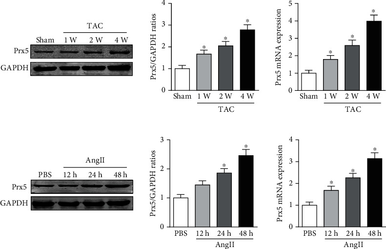
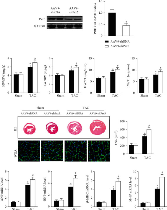
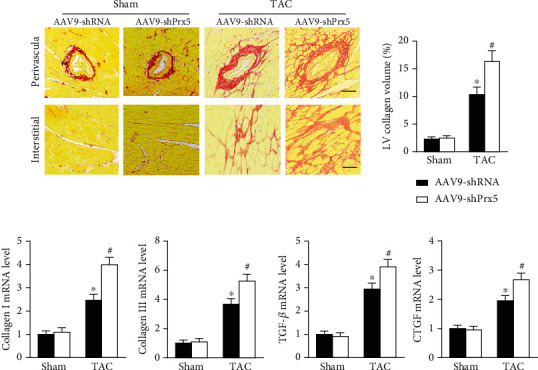
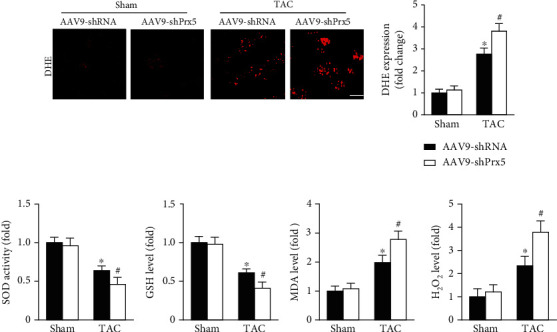
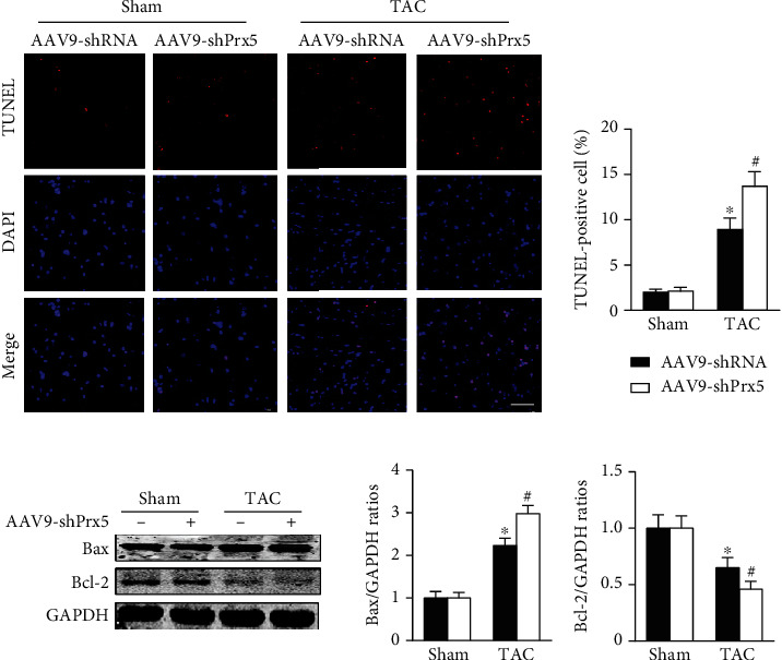
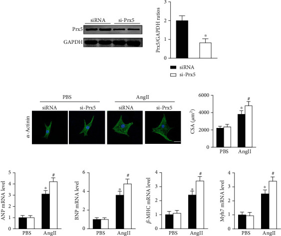
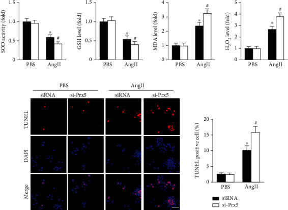
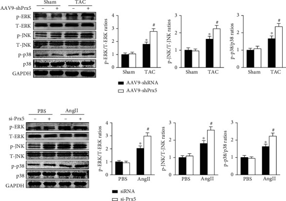
A recent study showed that peroxiredoxins (Prxs) play an important role in the development of pathological cardiac hypertrophy. However, the involvement of Prx5 in cardiac hypertrophy remains unclear. Therefore, this study is aimed at investigating the role and mechanisms of Prx5 in pathological cardiac hypertrophy and dysfunction. Transverse aortic constriction (TAC) surgery was performed to establish a pressure overload-induced cardiac hypertrophy model. In this study, we found that Prx5 expression was upregulated in hypertrophic hearts and cardiomyocytes. In addition, Prx5 knockdown accelerated pressure overload-induced cardiac hypertrophy and dysfunction in mice by activating oxidative stress and cardiomyocyte apoptosis. Importantly, heart deterioration caused by Prx5 knockdown was related to mitogen-activated protein kinase (MAPK) pathway activation. These findings suggest that Prx5 could be a novel target for treating cardiac hypertrophy and heart failure.
Copyright © 2022 Chengyun Hu et al.
Conflict of interest statement
No conflicts of interest are declared by the authors.
Figures
Similar articles
-
Mixed lineage kinase-3 prevents cardiac dysfunction and structural remodeling with pressure overload.Am J Physiol Heart Circ Physiol. 2019 Jan 1;316(1):H145-H159. doi: 10.1152/ajpheart.00029.2018. Epub 2018 Oct 26. Am J Physiol Heart Circ Physiol. 2019. PMID: 30362822 Free PMC article.
-
Peroxiredoxin-1 ameliorates pressure overload-induced cardiac hypertrophy and fibrosis.Biomed Pharmacother. 2020 Sep;129:110357. doi: 10.1016/j.biopha.2020.110357. Epub 2020 Jun 9. Biomed Pharmacother. 2020. PMID: 32531679
-
Hypoxia-Induced Mitogenic Factor Promotes Cardiac Hypertrophy via Calcium-Dependent and Hypoxia-Inducible Factor-1α Mechanisms.Hypertension. 2018 Aug;72(2):331-342. doi: 10.1161/HYPERTENSIONAHA.118.10845. Epub 2018 Jun 11. Hypertension. 2018. PMID: 29891648
-
MEF2 in cardiac hypertrophy in response to hypertension.Trends Cardiovasc Med. 2023 May;33(4):204-212. doi: 10.1016/j.tcm.2022.01.002. Epub 2022 Jan 11. Trends Cardiovasc Med. 2023. PMID: 35026393 Review.
-
Actin-Binding Proteins in Cardiac Hypertrophy.Cells. 2022 Nov 11;11(22):3566. doi: 10.3390/cells11223566. Cells. 2022. PMID: 36428995 Free PMC article. Review.
Cited by
-
Traditional Chinese medicine as a protective strategy against chemotherapy-induced cardiotoxicity: An overview of the literature.J Tradit Complement Med. 2024 Jun 22;15(2):107-118. doi: 10.1016/j.jtcme.2024.06.010. eCollection 2025 Mar. J Tradit Complement Med. 2024. PMID: 40060147 Free PMC article. Review.
-
Guanxinning tablets improve myocardial hypertrophy by inhibiting the activation of MEK-ERK1/2 signaling pathway.J Appl Biomed. 2023 Sep;21(3):137-149. doi: 10.32725/jab.2023.014. Epub 2023 Sep 18. J Appl Biomed. 2023. PMID: 37747313
-
Selenoprotein P, Peroxiredoxin-5, Renalase and Selected Cardiovascular Consequences Tested in Ambulatory Blood Pressure Monitoring and Echocardiography.Antioxidants (Basel). 2023 May 30;12(6):1187. doi: 10.3390/antiox12061187. Antioxidants (Basel). 2023. PMID: 37371917 Free PMC article.
References
-
- Mohammed S. A., Paramesha B., Kumar Y., Tariq U., Arava S. K., Banerjee S. K. Allylmethylsulfide, a sulfur compound derived from garlic, attenuates isoproterenol-induced cardiac hypertrophy in rats. Oxidative Medicine and Cellular Longevity . 2020;2020:15. doi: 10.1155/2020/7856318.7856318 - DOI - PMC - PubMed
MeSH terms
Substances
LinkOut - more resources
Full Text Sources
