

Levosimendan Postconditioning Attenuates Cardiomyocyte Apoptosis after Myocardial Infarction
- PMID: 35132355
- PMCID: PMC8817859
- DOI: 10.1155/2022/2988756
Levosimendan Postconditioning Attenuates Cardiomyocyte Apoptosis after Myocardial Infarction
Retraction in
-
Retracted: Levosimendan Postconditioning Attenuates Cardiomyocyte Apoptosis after Myocardial Infarction.J Healthc Eng. 2023 Aug 9;2023:9758474. doi: 10.1155/2023/9758474. eCollection 2023. J Healthc Eng. 2023. PMID: 37593472 Free PMC article.
Abstract
Background: Levosimendan preconditioning has been shown to attenuate myocardial apoptosis in animal models. However, protective effects of levosimendan postconditioning against myocardial apoptosis following myocardial infarction (MI) have not been evaluated. Therefore, we investigated the effects of levosimendan postconditioning on myocardial apoptosis in MI rat models.
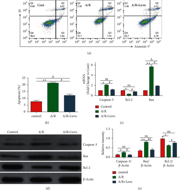
Methods: In an anoxia/reoxygenation (A/R) model, H9c2 cells were pretreated with or without levosimendan postconditioning after which their apoptosis rates were assessed by flow cytometry, RT-qPCR, and western blot analyses. Then, postconditioning was performed with or without levosimendan in MI rat models. Myocardiocyte apoptosis was evaluated by echocardiography, TTC staining, TUNEL staining, immunohistochemical staining, RT-qPCR, and western blot analysis.
Results: Levosimendan postconditioning inhibited H9c2 cell apoptosis in A/R models by elevating Bcl-2 while suppressing Caspase-3 and Bax at both mRNA and protein levels. Moreover, it improved cardiac functions and reduced the left ventricle infarction area in MI rat models. Compared to the MI control group, cardiomyocyte apoptosis rates in the levosimendan postconditioning group were low. The reduced cardiomyocyte apoptosis rates were associated with downregulation of Bax and Caspase-3 as well as with upregulation of Bcl-2 at mRNA and protein levels.
Conclusions: Levosimendan postconditioning of MI rat models protected against cardiomyocyte apoptosis, implying that it is a potential strategy for preventing cardiomyocyte apoptosis in the treatment of cardiac dysfunction following MI.
Copyright © 2022 Ying Xie et al.
Conflict of interest statement
The authors declare that there are no conflicts of interest regarding the publication of this paper.
Figures

References
-
- Dehmer G. J., Badhwar V., Bermudez E. A., et al. 2020AHA/ACC key data elements and definitions for coronary revascularization: a report of the American college of cardiology/American heart association task force on clinical data standards (writing committee to develop clinical data standards for coronary revascularization) Journal of the American College of Cardiology . 2021;75(16):1975–2088. doi: 10.1016/j.jacc.2020.02.010. - DOI - PubMed
-
- Visseren F. J., Mach F., Smulders Y. M. 2021 ESC Guidelines on cardiovascular disease prevention in clinical practice: developed by the Task Force for cardiovascular disease prevention in clinical practice with representatives of the European Society of Cardiology and 12 medical societies with the special contribution of the European Association of Preventive Cardiology (EAPC) European Heart Journal . 2021;42(34):3227–3337. doi: 10.1093/eurheartj/ehab484. - DOI - PubMed
Publication types
MeSH terms
Substances
LinkOut - more resources
Full Text Sources
Medical
Research Materials
