. 2022 Jun;18(2):223-239.
doi: 10.1007/s11302-022-09842-w. Epub 2022 Feb 7.

Optimized HPLC method to elucidate the complex purinergic signaling dynamics that regulate ATP, ADP, AMP, and adenosine levels in human blood

Affiliations

Optimized HPLC method to elucidate the complex purinergic signaling dynamics that regulate ATP, ADP, AMP, and adenosine levels in human blood

Carola Ledderose et al. Purinergic Signal. 2022 Jun.

Abstract

ATP released into the bloodstream regulates immune responses and other physiological functions. Excessive accumulation of extracellular ATP interferes with these functions, and elevated plasma ATP levels could indicate infections and other pathological disorders. However, there is considerable disagreement about what constitutes normal plasma ATP levels. Therefore, we optimized a method to accurately assess ATP concentrations in blood. We found that rapid chilling of heparinized blood samples is essential to preserve in vivo ATP levels and that differential centrifugation minimizes inadvertent ATP release due to cell damage and mechanical stress. Plasma samples were stabilized with perchloric acid, etheno-derivatized, and delipidated for sensitive analysis of ATP and related compounds using high-performance liquid chromatography (HPLC) and fluorescence detection. We measured 33 ± 20 nM ATP, 90 ± 45 nM ADP, 100 ± 55 nM AMP, and 81 ± 51 nM adenosine in the blood of healthy human adults (n = 10). In critically ill patients, ATP levels were 6 times higher than in healthy subjects. The anticoagulant greatly affected results. ATP levels were nearly 8 times higher in EDTA plasma than in heparin plasma, while AMP levels were 3 times lower and adenosine was entirely absent in EDTA plasma. If EDTA blood was not immediately chilled, ATP, ADP, and AMP levels continued to rise, which indicates that EDTA interferes with the endogenous mechanisms that regulate plasma adenylate levels. Our optimized method eliminates artifacts that prevent accurate determination of plasma adenylates and will be useful for studying mechanisms that regulate adenylate levels and for monitoring of pathological processes in patients with infections and other diseases.

Keywords: ATP; Adenosine; HPLC; Plasma adenylates; Purinergic signaling.


Conflict of interest statement

Carola Ledderose declares that she has no conflict of interest.

Eleftheria-Angeliki Valsami declares that she has no conflict of interest.

Wolfgang G. Junger declares that he has no conflict of interest.

Figures
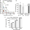
Fig. 1
Fig. 1
Rapid chilling of blood samples prevents ATP breakdown and adenosine uptake. a ATP (10 µM) was added to heparinized blood from a healthy human subject, and the blood was kept at 37 °C. After the indicated times, samples were chilled on ice, and plasma levels of ATP, ADP, AMP, and adenosine (ADO) were analyzed with HPLC. Data are representative of 3 experiments with blood from different healthy donors. b Chilling blood prevents ATP breakdown. ATP (10 µM) was added to heparinized human blood that was immediately chilled and processed for HPLC analysis or kept for 30 min either on ice or at 37 °C before processing. Results show means ± SD from 3 experiments with blood from different healthy donors. c Rapid chilling and processing of heparinized blood samples minimizes ATP breakdown. ATP at the indicated concentrations was added to chilled blood (n = 3 healthy donors) or kept in aqueous solution and analyzed by HPLC. Results show means ± SD
Fig. 2
Fig. 2
Streamlined process of plasma preparation preserves in vivo adenylate concentrations. Heparinized blood samples must be immediately chilled and kept on ice until they can be transferred to the research laboratory where blood cells and platelets are removed using a two-step centrifugation procedure. Resulting plasma samples are then treated with perchloric acid (PCA) at a final concentration of 400 mM in order to denature proteins and inactivate enzymes that can alter concentrations of ATP and its breakdown products. Samples stabilized in that manner can be frozen for long-term storage or shipping
Fig. 3
Fig. 3
Two-step centrifugation and PCA precipitation preserve in vivo adenylate levels. a Differential centrifugation prevents mechanically induced in vitro ATP release. Chilled heparinized blood samples (0 °C) from healthy human subjects were subjected to a single high-speed (2300 × g for 15 min) or a dual centrifugation procedure consisting of a low-speed (400 × g for 10 min) and a brief high-speed (2300 × g for 5 min) centrifugation step. Then, ATP, ADP, AMP, and adenosine (ADO) concentrations were analyzed by HPLC. Data are expressed as means ± SD of independent experiments with blood from different donors (n = 5); *p < 0.05 (t test). b Stabilizing plasma with perchloric acid (PCA) prevents the breakdown of ATP and its metabolites in vitro. Plasma samples of different healthy human subjects (n = 3) were deproteinized with PCA (400 mM) and spiked with 500 nM ATP. Samples were kept on ice and ATP, ADP, AMP, and adenosine concentrations were determined with HPLC at the indicated times (one-way ANOVA, p > 0.05)
Fig. 4
Fig. 4
Processing of plasma samples for etheno-derivatization of ATP and metabolites. PCA-treated plasma samples were defrosted in an ice bath and spiked with AMPCP as internal standard (final concentration of 250 nM). After removal of precipitated proteins by centrifugation, the supernatants were neutralized with K2HPO4 (0.4 M). After another centrifugation to remove remaining precipitates, the pH was adjusted to 5, and samples were treated with chloroacetaldehyde (C2H3ClO; 158.4 mM) and incubated for 30 min at 72 °C to generate etheno-derivatives. Samples were neutralized by addition of NH4HCO3 (100 mM)
Fig. 5
Fig. 5
Solid-phase extraction (SPE) with C18 columns to enrich adenylates and remove lipids. PCA-treated plasma samples processed as described in Fig. 4 to generate etheno-derivatives of ATP, ADP, AMP, and adenosine were concentrated by C18 solid-phase extraction (SPE). Samples mixed with HPLC buffer A containing the ion-pairing reagent tetrabutylammonium bisulfate were applied to the SPE columns. Then, the columns were rinsed twice with HPLC buffer A to eliminate small molecular contaminants. Finally, adenylates were eluted using HPLC buffer B. SPE columns containing poorly soluble lipids were discarded, and the eluents containing compounds of interest were dried using SpeedVac centrifugation. The dried samples were dissolved with KOH and HPLC buffer A. Insoluble material was spun down, discarded, and the supernatants filtered and subjected to HPLC analysis
Fig. 6
Fig. 6
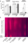
Solid-phase extraction with C18 columns effectively eliminates lipids without skewing levels of ATP and its metabolites. a PCA-treated plasma samples containing equal concentrations of ATP, ADP, AMP, adenosine (ADO), and AMPCP (1 µM, each) were subjected to C18 solid-phase extraction (SPE), chloroform/methanol (2:1 v/v; Folch method), or methanol (MeOH; 80%) extraction, analyzed by HPLC, and recovered adenylate concentrations were compared. Data are expressed as means ± SD of 3 separate experiments. b Equal amounts of plasma, PCA-treated plasma samples, and PCA-treated plasma samples that had been depleted from lipids by C18 SPE, the Folch extraction method, or by extraction with 80% methanol were separated by thin-layer chromatography. Lipids were visualized with Nile red staining. Results are representative of three experiments with similar results
Fig. 7
Fig. 7
ATP breakdown is attenuated in EDTA-treated blood samples. a Venous blood was drawn into pre-chilled EDTA or lithium heparin vacutainer tubes and ATP, ADP, AMP, and adenosine (ADO) concentrations were determined by HPLC. Data shown are means ± SD of independent experiments with blood from 10 different healthy donors; ***p < 0.001, t test. b Blood cells continue to release and metabolize ATP when samples are not immediately chilled. Blood samples were anticoagulated with EDTA or heparin and kept at room temperature. At the indicated times, plasma samples were processed and ATP, ADP, and AMP, and adenosine concentrations were measured by HPLC. Data show representative results of five experiments
Fig. 8
Fig. 8
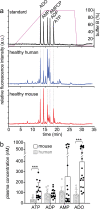
The optimized HPLC method is suitable for analysis of adenylate levels in mouse and human blood. Plasma samples from healthy C57BL/6 mice or from healthy human subjects were processed and analyzed by HPLC. a Representative chromatograms of HPLC standard, human plasma, and mouse plasma samples are shown. b Plasma ATP, ADP, AMP, and adenosine (ADO) concentrations differ markedly between human and murine subjects. Heparin blood samples from healthy human subjects (n = 10; age: 42 ± 9 years; data from Fig. 7) or healthy mice (n = 20, age 25.6 ± 8 weeks) were prepared and analyzed using the optimized HPLC assay. Data are shown as means ± SD, *p < 0.001, Mann–Whitney test
Fig. 9
Fig. 9
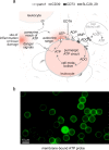
ATP release, breakdown, adenosine uptake, and metabolic recycling regulate pericellular ATP levels in human blood. a Model by which pericellular and global ATP influence immune cell function. Extracellular ATP concentrations depend on ATP released from inflamed or damaged tissues and the pericellular ATP release from blood cells via pannexin 1 (panx1). ATP levels are also influenced by dilution in the extracellular space and by ectoenzymes such as CD39 and CD73 that generate ADP, AMP, and adenosine, which is returned for cellular recycling via concentrative (CNT) and equilibrative (ENT) nucleoside transporters (SLC28 and SLC29). Optimized methods for blood sample processing must minimize interference with these processes in order to avoid skewing of actual in vivo adenylate levels. b Local pericellular ATP levels are restricted to subcellular sites that regulate immune cell functions. Human neutrophils labeled with a membrane-bound fluorescence ATP probe [24] reveal local hotspots of ATP in the immediate pericellular environment

Similar articles

Cited by

References

    1. Khakh BS, Burnstock G. The double life of ATP. Sci Am. 2009;301(84–90):92. doi: 10.1038/scientificamerican1209-84. - DOI - PMC - PubMed
    1. Burnstock G. Physiology and pathophysiology of purinergic neurotransmission. Physiol Rev. 2007;87:659–797. doi: 10.1152/physrev.00043.2006. - DOI - PubMed
    1. Ralevic V, Burnstock G. Receptors for purines and pyrimidines. Pharmacol Rev. 1998;50:413–492. - PubMed
    1. Antonioli L, Blandizzi C, Pacher P, Haskó G. The purinergic system as a pharmacological target for the treatment of immune-mediated inflammatory diseases. Pharmacol Rev. 2019;71:345–382. doi: 10.1124/pr.117.014878. - DOI - PMC - PubMed
    1. Corriden R, Insel PA (2010) Basal release of ATP: an autocrine-paracrine mechanism for cell regulation. Sci Signal 3:re1. 10.1126/scisignal.3104re1 - PMC - PubMed

Publication types

LinkOut - more resources