. 2022 Feb 15;11(4):e022070.
doi: 10.1161/JAHA.121.022070. Epub 2022 Feb 8.

Myogenic Vasoconstriction Requires Canonical Gq/11 Signaling of the Angiotensin II Type 1 Receptor

Affiliations

Myogenic Vasoconstriction Requires Canonical Gq/11 Signaling of the Angiotensin II Type 1 Receptor

Yingqiu Cui et al. J Am Heart Assoc. .

Abstract

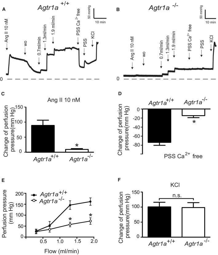
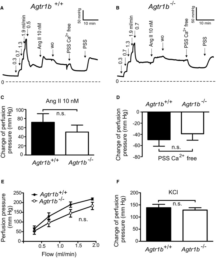
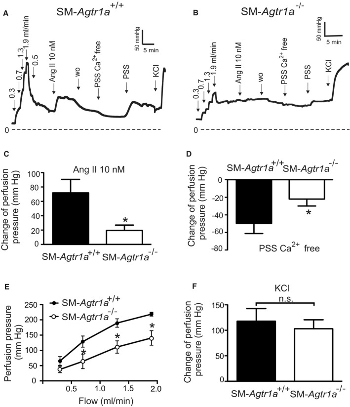
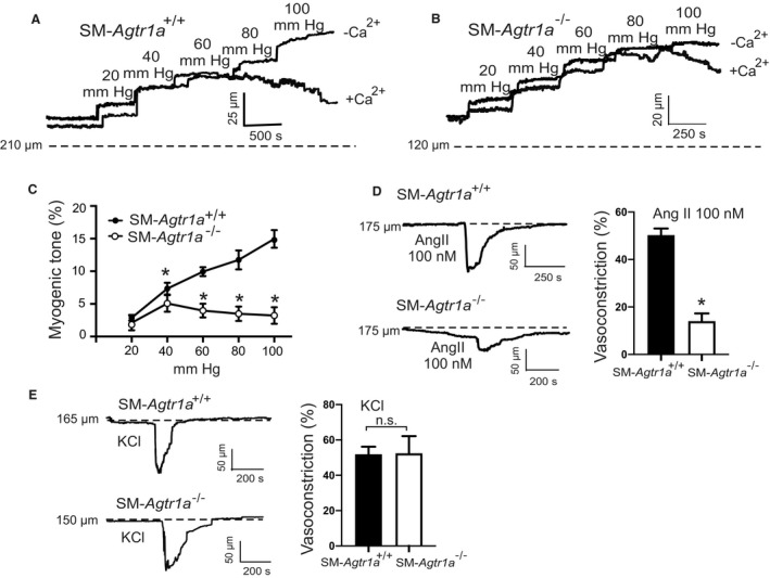
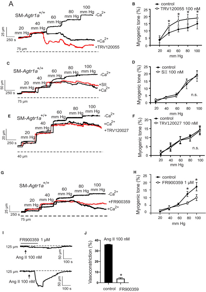
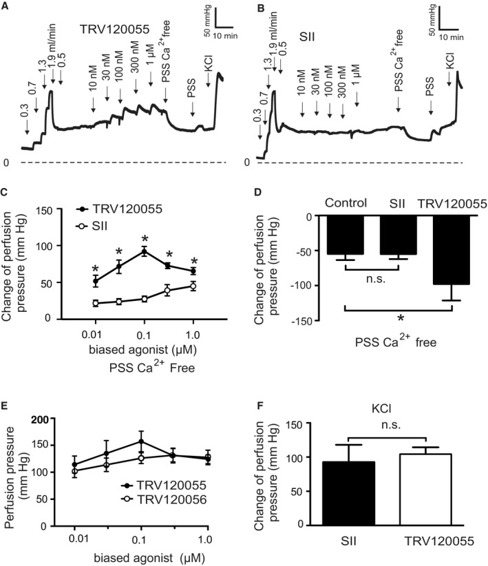
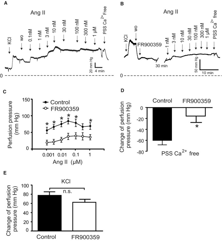
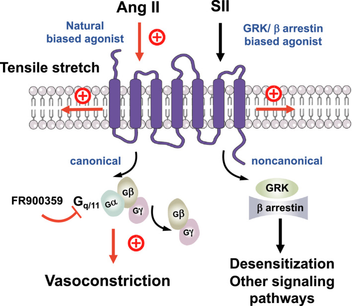
Background Blood pressure and tissue perfusion are controlled in part by the level of intrinsic (myogenic) arterial tone. However, many of the molecular determinants of this response are unknown. We previously found that mice with targeted disruption of the gene encoding the angiotensin II type 1a receptor (AT1AR) (Agtr1a), the major murine angiotensin II type 1 receptor (AT1R) isoform, showed reduced myogenic tone; however, uncontrolled genetic events (in this case, gene ablation) can lead to phenotypes that are difficult or impossible to interpret. Methods and Results We tested the mechanosensitive function of AT1R using tamoxifen-inducible smooth muscle-specific AT1aR knockout (smooth muscle-Agtr1a-/-) mice and studied downstream signaling cascades mediated by Gq/11 and/or β-arrestins. FR900359, Sar1Ile4Ile8-angiotensin II (SII), TRV120027 and TRV120055 were used as selective Gq/11 inhibitor and biased agonists to activate noncanonical β-arrestin and canonical Gq/11 signaling of the AT1R, respectively. Myogenic and Ang II-induced constrictions were diminished in the perfused renal vasculature, mesenteric and cerebral arteries of smooth muscle-Agtr1a-/- mice. Similar effects were observed in arteries of global mutant Agtr1a-/- but not Agtr1b-/- mice. FR900359 decreased myogenic tone and angiotensin II-induced constrictions whereas selective biased targeting of AT1R-β-arrestin signaling pathways had no effects. Conclusions This study demonstrates that myogenic arterial constriction requires Gq/11-dependent signaling pathways of mechanoactivated AT1R but not G protein-independent, noncanonical pathways in smooth muscle cells.

Keywords: angiotensin II type 1a receptor; arterial smooth muscle; biased ligands; myogenic vasoconstriction.


Figures

Figure 1
Figure 1. Vasoregulation in isolated perfused kidneys of Agtr1a −/− mice.
A and B, Original recordings of perfusion pressure in kidneys of Agtr1a +/+ (A) and Agtr1a −/− mice (B). C, Increase in the perfusion pressure induced by 10 nmol/L Ang II. D, Myogenic tone assessed by exposure to Ca2+ free PSS. E, Perfusion pressure at flow rates of 0.3, 0.7, 1.3, and 1.9 mL/min. F, Increase in the perfusion pressure induced by 60 mmol/L KCl. n=6 Agtr1a +/+ kidneys from 6 mice and n=7 Agtr1a −/− kidneys from 7 mice for all panels. *P<0.05. Ang II indicates angiotensin II; n.s., not significant; PSS, physiological saline solution; and wo, wash‐out.
Figure 2
Figure 2. Vasoregulation in isolated perfused kidneys of Agtr1b −/− mice.
A and B, Original recordings of the perfusion pressure in kidneys of Agtr1b +/+ (A) and Agtr1b −/− mice (B). C, Increase in perfusion pressure induced by 10 nmol/L Ang II. D, Change of pressure assessed by exposure to Ca2+ free PSS. E, Perfusion pressure at flow rates of 0.3, 0.7, 1.3, and 1.9 mL/min. F, Increase in perfusion pressure induced by 60 mmol/L KCl. n=6 Agtr1b +/+ kidneys from 5 mice and n=6 Agtr1b −/− kidneys from 3 mice for all panels. Ang II indicates angiotensin II; n.s., not significant; PSS, physiological saline solution; and w.o., washout.
Figure 3
Figure 3. Vasoregulation in isolated perfused kidneys of SM‐Agtr1a −/− mice.
A and B, Original recordings of the perfusion pressure in kidneys of SM‐Agtr1a +/+ (A) and SM‐Agtr1a −/− mice (B). C, Increase in perfusion pressure induced by 10 nmol/L Ang II. D, Change of pressure assessed by exposure to Ca2+ free PSS. E, Perfusion pressure at flow rates of 0.3, 0.7, 1.3, and 1.9 mL/min. F, Increase in perfusion pressure induced by 60 mmol/L KCl. n=6 SM‐Agtr1a +/+ kidneys from 5 mice and n=6 SM‐Agtr1a −/− kidneys from 6 mice for all panels. *P<0.05. Ang II indicates angiotensin II; n.s., not significant; PSS, physiological saline solution; SM, smooth muscle; and w.o., washout.
Figure 4
Figure 4. Myogenic tone in mesenteric arteries.
A and B, Representative recordings of MA diameter during a series of pressure steps from 20 to 100 mm Hg in 20 mm Hg increments in control conditions (+Ca2+) and in Ca2+ free solution (−Ca2+). Arteries were isolated from SM‐Agtr1a +/+ (A) and SM‐Agtr1a −/− mice (B). Note the increase in active constriction over the entire pressure range from 60 to 100 mm Hg in vessels from SM‐Agtr1a +/+, but not from SM‐Agtr1a −/− mice. Vasodilation in Ca2+‐free solution was observed in SM‐Agtr1a +/+ but not in SM‐Agtr1a −/− arteries (P<0.05). C, Average myogenic tone of mesenteric arteries in PSS expressed as dilation of vessels induced by external Ca2+ free solution (0 Ca/EGTA; SM‐Agtr1a +/+, n=9, and SM‐Agtr1a −/−, n=6 vessels, each from individual mice for both groups). D, Response to Ang II and (E) response to 60 mmol/L KCl in MA of SM‐Agtr1a +/+ and SM‐Agtr1a −/− mice. MAs were pressurized to 80 mm Hg. Responses are expressed as relative changes in vessel inner diameter. SM‐Agtr1a +/+, n=5 vessels, and SM‐Agtr1a −/−, n=4 vessels, from 5 and 4 mice, respectively. *P<0.05. Ang II indicates angiotensin II; MA, mesenteric arteries; PSS, physiological saline solution; and SM, smooth muscle.
Figure 5
Figure 5. Enhancement of the vascular tone by TRV120055.
A, C, E, and G, Representative recordings of mesenteric artery diameter during a series of pressure steps from 20 to 100 mm Hg in 20 mm Hg increments in control conditions (+Ca2+), TRV120055 100 nmol/L (A), SII 100 nmol/L (C), TRV120027 100 nmol/L (E), FR900359 1 µmol/L (G) and in Ca2+‐free solution. B, D, F, and H, Average myogenic constriction of mesenteric arteries in drug‐free physiological saline solution and in PSS containing 100 nmol/L TRV120055 (B), 100 nmol/L SII (D), TRV120027 100 nmol/L (F) and 1 µmol/L FR900359 (H) (n=6, 4, 5 and 4, respectively, each from individual mice). I and J, Response to Ang II in MA in drug‐free PSS and PSS in presence of FR900359 at 80 mm Hg (n=6 each from individual mice). *P<0.05. Ang II indicates angiotensin II; n.s. indicates not significant; and SII, Sar1Ile4Ile8‐angiotensin.
Figure 6
Figure 6. Function of biased AT1R agonists to vasoregulation in isolated perfused kidneys from SM‐Agtr1a +/+ mice.
A and B, Original recordings of perfusion pressure in response to various flow rates (in mL/min), TRV120055 (A) or Sar1Ile4Ile8‐SII (SII) (B), Ca2+ free perfusion solution (PSS Ca2+ free) and reexposure of the kidneys to PSS. C, Increase in perfusion pressure induced by TRV120055 and SII in various concentrations (10 nmol/L to 1 µmol/L). D, Change of perfusion pressure assessed by exposure of the kidneys to Ca2+ free PSS at the presence of TRV120055 or Sar‐Ile II at the concentration of 100 nmol/L. E, Dose‐response relationships for TRV120055 and TRV120056. F, Increase in perfusion pressure induced by 60 mmol/L KCl. TRV120055, TRV120056, SII. n=6 kidneys from 5 mice in each group; n=6 kidneys from 5 mice in the control group. *P<0.05. AT1R, angiotensin II type 1 receptor; Control, SM‐Agtr1a +/+ without biased ligand; n.s., not significant; PSS, physiological saline solution; and SM, smooth muscle.
Figure 7
Figure 7. Vasoregulation in isolated perfused kidneys of SM‐Agtr1a +/+ mice pretreated with 300 nmol/L Gq/11 blocker FR900359.
A, Original recordings of perfusion pressure in kidneys of Agtr +/+ mice in response to various concentrations of Ang II (B) same as (A) but pretreated with 300 nmol/L FR900359 for 30 minutes. C, Increases in perfusion pressure induced by Ang II (1 nmol/L to 1 µmol/L). D, Myogenic tone assessed by exposure of the kidneys to Ca2+‐free PSS. E, Increase in perfusion pressure induced by 60 mmol/L KCl. n=15 SM‐Agtr1a +/+ kidneys from 12 mice and n=6 SM‐Agtr1a +/+ kidneys from 4 mice pretreated with FR900359 for all panels. *P<0.05. Ang II indicates angiotensin II; n.s., not significant; PSS, physiological saline solution; SM, smooth muscle; and w.o., wash‐out.
Figure 8
Figure 8. Schematic illustration of angiotensin II type 1a receptor (AT1aR) biased signaling cascade regulating myogenic arterial tone.
Canonical Gq/11 signaling pathway of the AT1R (purple blue) causes myogenic vasoconstriction whereas noncanonical β‐arrestin‐biased signaling is not involved in this process. Gq/11 proteins are heterotrimeric G proteins, which are made up of alpha (α), beta (β), and gamma (γ) subunits. The alpha subunit is attached to either a guanosine triphosphate (GTP) or guanosine diphosphate (GDP), which serves as an on‐off switch for the activation of the G‐protein. Upon activation of the AT1aR by either ligand‐independent mechanical stretch or the natural‐biased ligand Ang II, the Gβγ complex is released from the Gα subunit after its GDP‐GTP exchange for canonical G protein signaling to cause myogenic and/or humoral (Ang II‐mediated) vasoconstriction. This pathway is inhibited by the Gq/11 inhibitor FR900359. Although, GRKs and arrestins play a role in multiple noncanonical signaling pathways in cells, this pathway is unlikely engaged by mechanoactivated AT1Rs in response to tensile stretch or their natural ligand angiotensin II to cause vasoconstriction. Ang II indicates angiotensin II; GRK, G protein‐coupled receptor kinase; and SII, Sar1Ile4Ile8‐angiotensin.

Comment in

References

    1. Bayliss WM. On the local reactions of the arterial wall to changes of internal pressure. J Physiol. 1902;28:220–231. doi: 10.1113/jphysiol.1902.sp000911 - DOI - PMC - PubMed
    1. Davis MJ. Perspective: physiological role(s) of the vascular myogenic response. Microcirculation. 2012;19:99–114. doi: 10.1111/j.1549-8719.2011.00131.x - DOI - PubMed
    1. Cipolla MJ, Curry AB. Middle cerebral artery function after stroke: the threshold duration of reperfusion for myogenic activity. Stroke. 2002;33:2094–2099. doi: 10.1161/01.STR.0000020712.84444.8D - DOI - PubMed
    1. Gschwend S, Henning RH, Pinto YM, de Zeeuw D, van Gilst WH, Buikema H. Myogenic constriction is increased in mesenteric resistance arteries from rats with chronic heart failure: instantaneous counteraction by acute AT1 receptor blockade. Br J Pharmacol. 2003;139:1317–1325. - PMC - PubMed
    1. Ledoux J, Gee DM, Leblanc N. Increased peripheral resistance in heart failure: new evidence suggests an alteration in vascular smooth muscle function. Br J Pharmacol. 2003;139:1245–1248. doi: 10.1038/sj.bjp.0705366 - DOI - PMC - PubMed

Publication types

MeSH terms

LinkOut - more resources