. 2022 Feb 8;7(3):e153913.
doi: 10.1172/jci.insight.153913.

High-resolution structure-function mapping of intact hearts reveals altered sympathetic control of infarct border zones

Affiliations

High-resolution structure-function mapping of intact hearts reveals altered sympathetic control of infarct border zones

Ching Zhu et al. JCI Insight. .

Abstract

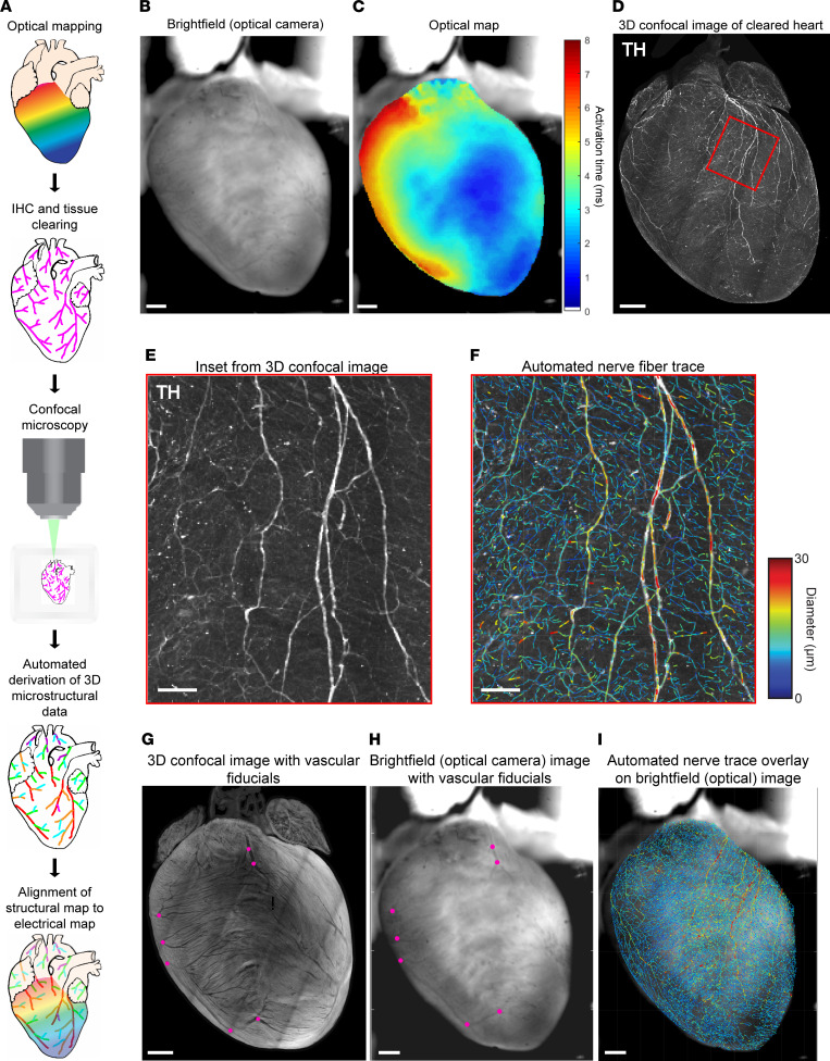
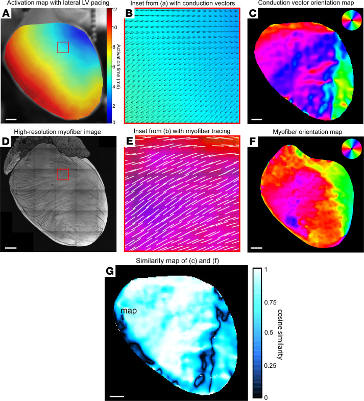
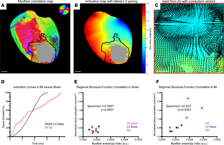
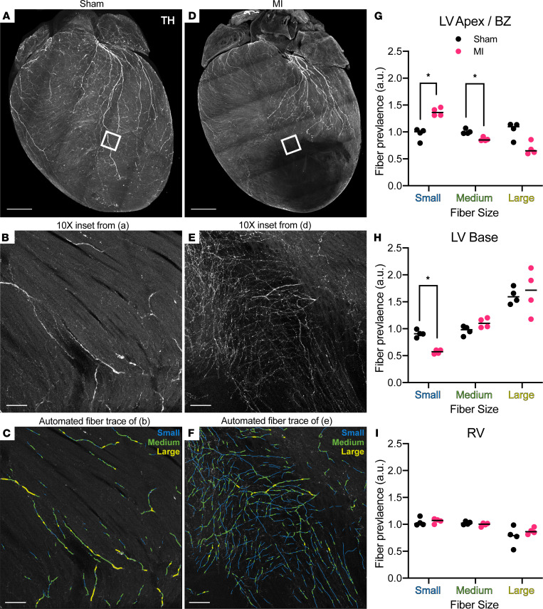
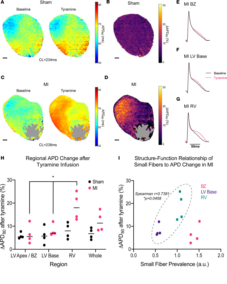
Remodeling of injured sympathetic nerves on the heart after myocardial infarction (MI) contributes to adverse outcomes such as sudden arrhythmic death, yet the underlying structural mechanisms are poorly understood. We sought to examine microstructural changes on the heart after MI and to directly link these changes with electrical dysfunction. We developed a high-resolution pipeline for anatomically precise alignment of electrical maps with structural myofiber and nerve-fiber maps created by customized computer vision algorithms. Using this integrative approach in a mouse model, we identified distinct structure-function correlates to objectively delineate the infarct border zone, a known source of arrhythmias after MI. During tyramine-induced sympathetic nerve activation, we demonstrated regional patterns of altered electrical conduction aligned directly with altered neuroeffector junction distribution, pointing to potential neural substrates for cardiac arrhythmia. This study establishes a synergistic framework for examining structure-function relationships after MI with microscopic precision that has potential to advance understanding of arrhythmogenic mechanisms.

Keywords: Arrhythmias; Cardiology; Innervation; Mouse models.


Conflict of interest statement

Conflict of interest: KS and PSR are cofounders of and have direct ownership in NeuTrace Inc. and NeuFera Inc. PSR receives income from NeuTrace Inc. KS and PSR are inventors of intellectual property pertaining to cardiac neural diagnostics and therapeutics.

Figures

Figure 1
Figure 1. Optical mapping and tissue clearing pipeline to align electrical and structural maps.
(A) Schematic of optical mapping, clearing, imaging, and automated feature tracing steps in the alignment pipeline. (B and C) Bright-field image taken simultaneously with optical action potential map showing activation in sinus rhythm. (D) Maximum intensity projection (MIP) image of tyrosine hydroxylase–positive (TH-positive) nerve fibers on the ventral surface of the same heart after IHC, tissue clearing, and confocal imaging. (E and F) High-magnification images of the boxed region in D, with TH staining alongside nerve-fiber tracing by computer vision, color-coded by fiber diameter. (G and H) Venous bifurcations (magenta points) on MIP confocal shell image of a cleared heart alongside bright-field image of same heart were used as fiducial anchors for alignment. (I) Automated global nerve-fiber tracing aligned with bright-field image allows spatial correlation with optical action potential data. Scale bars: 1 mm (B–D and G–I); 100 μm (E and F).
Figure 2
Figure 2. Structure-function alignment correlates global ventricular impulse propagation with myofiber orientation.
(A) Optical activation map of representative sham heart with basolateral left ventricular (LV) pacing. (B) High-magnification image of the boxed region in A showing an activation map with overlay of conduction velocity vectors calculated in ElectroMap. (C) Global ventricular conduction vector orientation map color-coded by vector angle. (D) Maximum intensity projection confocal image of muscle autofluorescence with high-resolution myofiber structure. (E) High-magnification image of the boxed region in D, with overlay of automated myofiber orientation tracing. (F) Global ventricular myofiber orientation map color-coded by fiber angle. (G) Cosine similarity map calculated by taking cosine of angular difference between C and F. Scale bars: 1 mm (A, C, D, F, and G).
Figure 3
Figure 3. A composite metric of myofiber anisotropy and tissue activation defines infarct border zones.
(A) Color-coded myofiber orientation map from chronic myocardial infarction (MI) heart with black line delineating border zone (BZ) and gray patch delineating dense scar. Dark patch on left ventricular (LV) lateral wall denotes location of coronary ligature, which was excluded from quantitative analyses. Atria were cropped from image for ease of interpretation. (B) Activation map from same chronic MI heart with dense scar region defined by gray patch and BZ delineated by black line. (C) High-magnification image of the boxed region in B, with activation map with overlay of conduction velocity vectors showing discontinuous propagation. (D) Representative tissue activation curves from anatomically defined LV apex region of sham heart (black) versus infarct BZ region (magenta), showing isotropic conduction versus anisotropic and conduction block. (E and F) Plots of regional myofiber anisotropy indices versus normalized tissue activation times, showing no correlation in sham (Spearman’s r = 0.0667, P = 0.8801, n = 9 regions from 3 mice) versus positive correlation in MI (Spearman’s r = 0.833, P = 0.0083, n = 9 regions from 3 mice). Scale bars: 1 mm (A and B); 100 μm (C).
Figure 4
Figure 4. Altered distribution of neuroeffector endings after myocardial infarction.
(A) Maximum intensity projection (MIP) confocal image of global tyrosine hydroxylase (TH) staining of sham heart (same heart MIP was originally shown in Figure 1D for methodological demonstration purposes). (B and C) Representative images from boxed region in A (original magnification, ×10) from sham left ventricle (LV) showing TH staining and automated fiber tracing, binned by small (1.2–3 μm), medium (3–5 μm), and large (5–100 μm) diameters. (D) MIP confocal image of global TH staining in myocardial infarction (MI) heart. (E and F) Representative images from boxed region in D (original magnification, ×10) from border zone (BZ) showing TH staining and automated fiber tracing, binned by small (1.2–3 μm), medium (3–5 μm), and large (5–100 μm) diameters. (G–I) Regional comparisons of fiber size prevalence between sham (black) and MI (magenta), with black lines denoting medians and asterisks denoting statistical significance (Mann-Whitney, *P = 0.0286, n = 4 mice per group). Scale bars: 1 mm (A and D); 100 μm (B, C, E, and F). RV, right ventricle.
Figure 5
Figure 5. Altered neuroeffector distribution underlies perturbed myocardial sympathetic control after chronic infarction.
(A) Eighty percent of action potential duration (APD80) maps of representative sham heart at baseline and after infusion of 5 μM tyramine. (B) Change in APD80 (ΔAPD80) map of sham heart. (C) APD80 maps of representative MI heart at baseline and after infusion of 5 μM tyramine. Gray region denotes dense scar. (D) APD80 map of MI heart. (E–G) Representative action potentials at baseline (black) and after tyramine (magenta) in anatomically segmented regions of MI heart. (H) Comparison of regional, tyramine-mediated changes in APD80 between sham and MI hearts, with MI hearts showing significant regional variation in tyramine effect (Kruskal-Wallis, *P = 0.0132, n = 4 mice per group) while sham hearts showed no significant regional variation (Kruskal Wallis, P = 0.7463, n = 4 mice per group). (I) Plot of regional small-fiber prevalence in MI hearts versus tyramine-mediated APD change, with positive correlation in left ventricular (LV) base and right ventricular (RV) regions (Spearman’s r = 0.7381, P = 0.0458, n = 8 regions from 4 mice) but no correlation when border zone (BZ) is included (Spearman’s r = 0.021, P = 0.956, n = 12 regions from 4 mice). Scale bars: 1 mm (A–D).

References

    1. Zipes DP, Wellens HJ. Sudden cardiac death. Circulation. 1998;98(21):2334–2351. doi: 10.1161/01.CIR.98.21.2334. - DOI - PubMed
    1. Chugh SS, et al. Epidemiology of sudden cardiac death: clinical and research implications. Prog Cardiovasc Dis. 2008;51(3):213–228. doi: 10.1016/j.pcad.2008.06.003. - DOI - PMC - PubMed
    1. El-Sherif N, et al. Re-entrant ventricular arrhythmias in the late myocardial infarction period. 1. Conduction characteristics in the infarction zone. Circulation. 1977;55(5):686–702. doi: 10.1161/01.CIR.55.5.686. - DOI - PubMed
    1. Peters NS, et al. Disturbed connexin43 gap junction distribution correlates with the location of reentrant circuits in the epicardial border zone of healing canine infarcts that cause ventricular tachycardia. Circulation. 1997;95(4):988–996. doi: 10.1161/01.CIR.95.4.988. - DOI - PubMed
    1. Kléber AG, Rudy Y. Basic mechanisms of cardiac impulse propagation and associated arrhythmias. Physiol Rev. 2004;84(2):431–488. doi: 10.1152/physrev.00025.2003. - DOI - PubMed

Publication types