Review
. 2022 Feb;42(2):88-116.
doi: 10.1002/cac2.12250.

Ferroptosis in cancer and cancer immunotherapy

Affiliations
Review

Ferroptosis in cancer and cancer immunotherapy

Lei Zhao et al. Cancer Commun (Lond). 2022 Feb.

Abstract

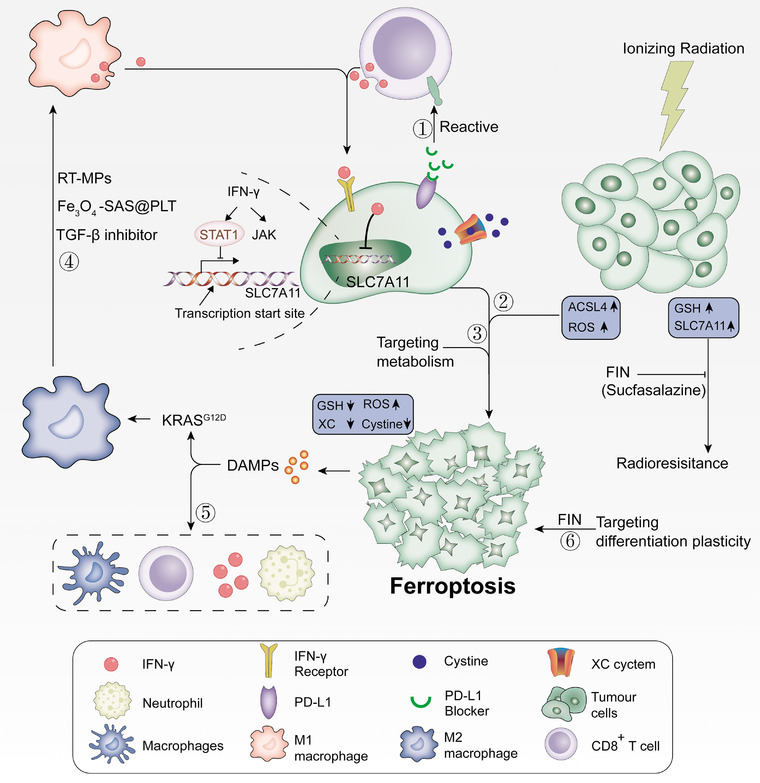
The hallmark of tumorigenesis is the successful circumvention of cell death regulation for achieving unlimited replication and immortality. Ferroptosis is a newly identified type of cell death dependent on lipid peroxidation which differs from classical programmed cell death in terms of morphology, physiology and biochemistry. The broad spectrum of injury and tumor tolerance are the main reasons for radiotherapy and chemotherapy failure. The effective rate of tumor immunotherapy as a new treatment method is less than 30%. Ferroptosis can be seen in radiotherapy, chemotherapy, and tumor immunotherapy; therefore, ferroptosis activation may be a potential strategy to overcome the drug resistance mechanism of traditional cancer treatments. In this review, the characteristics and causes of cell death by lipid peroxidation in ferroptosis are briefly described. In addition, the three metabolic regulations of ferroptosis and its crosstalk with classical signaling pathways are summarized. Collectively, these findings suggest the vital role of ferroptosis in immunotherapy based on the interaction of ferroptosis with tumor immunotherapy, chemotherapy and radiotherapy, thus, indicating the remarkable potential of ferroptosis in cancer treatment.

Keywords: cancer; ferroptosis; immunotherapy.


Conflict of interest statement

The authors declare that they have no competing interests.

Figures

FIGURE 1
FIGURE 1
The occurrence of ferroptosis. (A) Generation of OH· and O2·. Oxygen produces O2· under the action of NADPH or NOX and mitochondria. Subsequently, O2· is converted into H2O2 under the action of SOD. H2O2 forms HO· in the presence of iron ions, which deprive PUFA of hydrogen atoms, causing lipid peroxidation. (B) Lipid peroxidation cycle and its termination. HO· or RO· catalyzes R to form RO·, RO· reacts quickly with oxygen to form ROO·, then extracts hydrogen from another R to produce new RO· and ROOH (positive feedback chain reaction). GPX4 can be used as an antioxidant to reduce ROOH to R to stop the peroxidation reaction. (C) The occurrence of ferroptosis. ROO· will decompose to produce MDA and 4‐HNE, then form covalent adducts with macromolecules such as protein, DNA, and lipids, or crosslink and inactivate proteins that promote ferroptosis, thereby promoting cell membrane rupture and ferroptosis. Abbreviations: NOX, NADPH oxidase; O2, superoxide radicals; SOD, peroxidase; HO·, hydroxyl radicals; RO·, lipid free radicals; ROO·, lipid peroxy‐free radicals; ROOH, lipids hydroperoxide; MDA, malondialdehyde; POR, NADPH‐cytochrome P450 reductase; CYB5R1, NADH‐cytochrome b5 reductase; CYP450, cytochrome P450; NOX, NADPH oxidase; SOD, superoxide dismutase; GPX4, glutathione peroxidase 4; 4‐HNE, 4‐Hydroxy‐2‐nonanal
FIGURE 2
FIGURE 2
Ferroptosis metabolic pathway. (A) Antioxidant metabolism. GPX4 and CoQ10 are two parallel pathways of antioxidant metabolism. GPX4 exerts an antioxidant function through the metabolism of GSH. CoQ10 is reduced to CQ10H2 under the action of FSP1, inhibiting lipid peroxidation by capturing lipid peroxidation free radicals. (B) Iron metabolism. Iron binds to TF and is released into the cytoplasm by TFRC. Ferritin releases a large amount of iron‐by‐iron autophagy mediated by NACO4. Heme releases iron under the catalysis of HO‐1. FPN can transport iron from cells to the blood. (C) Lipid metabolism signaling pathway. AA/AdA is converted into polyunsaturated fatty acids (PE‐AA) by ACSL4 and LPCAT3. The balance between lipid synthesis, storage, and degradation can also manipulate the ferroptosis process. Acetyl‐CoA can also produce PUFA under the action of ACC synthase, immediately produce a large amount of ROS under the action of Fe2+ and LOX15. Abbreviations: GPX4, glutathione peroxidase 4; CoQ10, ubiquinone; FSP1, ferroptosis inhibitor protein 1; TF, transferrin; TFRC, transferrin receptor 1; HO‐1, heme oxygenase‐1; NFS1, cysteine desulfurase 1; ACSL4, acyl‐CoA synthetase 4; AA, arachidonic acid; AdA, epinephrine; LPCAT3, lysophosphatidylcholine acyltransferase 3; ATG5, autophagy‐related 5; BACO4, ###; BH4, tetrahydrobiopterin; Cys, Cysteine; DMT1, divalent metal transporter; FPN, ferroportin; GCLC, glutamate‐cysteine ligase catalytic subunit; Gln, Glutamine; Glu, Glutamate; GR, gluathione reductase; GSH, glutathione; GSS, Glutathione synthetase; GSSH, glutathione counterpart; iFSP1, Inhibit FSP1; iNFS1, Inhibit NFS1; LOX15, lipoxygenase 15; NADPH, nicotinamide adenine dinucleotide phosphate; NOX, NADPH oxidase; PE, Phosphatidylethanolamine; RAB7A, a small GTPase of the Rab family; SLC3A2, solute carrier family 3 member 2; SLC7A11, cystine/glutamate antiporter solute carrier family 7 member 11; STEAP3, Six‐transmembrane epithelial antigen of the prostate 3
FIGURE 3
FIGURE 3
Crosstalk between ferroptosis and classic signaling pathways. (A) AMPK‐ACC‐PUFA pathway. EMP, PPP and TCA cycles all require glucose as a substrate. When glucose is lacking, insufficient intracellular energy metabolism will activate AMPK, reducing the synthesis of PUFA by inhibiting ACC. (B) HCAR/MCT1‐SREBP1‐SCD1 pathway. Lactate enhances cell lipid synthesis by inhibiting glycolysis. Lactate induces the expression of HCAR1, MCT1 and inhibiting ferroptosis by directly inhibit ACSL4. Moreover, lactate promotes the phosphorylation of AMPK, which inhibits the ability of SREBP1 to promote SCD1, which directly inhibit ferroptosis by promoting the production of CQ10 and MUFA. (C) MTORC1‐Keap1‐NRF2 pathway. PI3K‐AKT‐mTORC1 signaling pathway activation will upregulate SREBP1 and SCD1, promoting MUFA production to resist ferroptosis. In addition to degrading SLC7A11, mTORC1 also promotes the accumulation of NRF2 by phosphorylation of p62, and indirectly regulates NRF2 target genes. mTORC2 directly regulates the phosphorylation of SLC7A11. (D) Cadherin‐NE2‐Hippo‐YAP pathway. At high cell density, ECAD will send a signal to the Hippo signaling pathway through NF2, inhibiting the ferroptosis process by promoting the phosphorylation of YAP and TZA. The green box indicates inhibition of ferroptosis, and the red box indicates the inducer of ferroptosis. Abbreviations: HCAR1, lactate receptor; MCT1, lactate transporter; SREBP1, sterol regulatory element‐binding protein 1; SCD1, stearoyl‐CoA desaturase 1; MUFA, monounsaturated fatty acid; ACC, acetyl‐coenzyme A carboxylase; ACSL4, acyl‐CoA synthetase long‐chain family member 4; AMP, adenosine monophosphate; AMPK, AMP‐activated protein kinase; ATP, adenosine triphosphate; BECN1, beclin 1; EMP, Embden‐Meyerhof‐Parnas pathway; FTH, ferritin heavy chain; GCL, glutamate cysteine ligase; GPX4, glutathione peroxidase 4; GSS, glutathione synthetase; LPCAT3, lysophosphatidylcholine acyltransferase 3; NF2, neurofibromatosis type 2; NOX4, NADPH oxidase 4; NRF2, NFE2‐related factor 2; PI3K, phosphatidylinositol 3‐kinase; PTEN, phosphatase and tensin homolog; PUFA, polyunsaturated fatty acids; SLC7A11, cystine/glutamate antiporter solute carrier family 7 member 11; SOD1, superoxide dismutase; TAZ, PDZ‐binding motif; TCA, tricarboxylic acid; TSC, tuberous sclerosis; YAP, yes‐associated protein
FIGURE 4
FIGURE 4
Ferroptosis and drug treatment of tumor. (A) Classic cancer and corresponding treatment drugs of ferroptosis. (B) Small molecules used in ferroptosis therapy to treat cancer
FIGURE 5
FIGURE 5
Ferroptosis and tumor immunotherapy. CD8+ T cells inhibit tumor cell cystine uptake by downregulating SLC3A2 and SLC7A11 by releasing IFNγ, and at the same time assisting immune checkpoint inhibitor PD‐L1, which can synergistically enhance T cell‐mediated anti‐tumor immunity and induce tumor cell ferroptosis. Ferroptosis is dependent on glucose and glutamine, and selectively targeting tumor cells by hindering metabolism has a bright future. The DAMPs released in vitro by cancer cells undergoing ferroptosis can induce the maturation of dendritic cells, cross‐induction of CD8+ T cells, production of IFN‐γ, and production of M2 macrophages. Subsequently, it activates adaptability in the tumor microenvironment, forming a positive feedback of the immune response. Radiotherapy produces a large amount of ROS by upregulating ACSL4 and inactivating SLC7A11 or GPX4 with ferroptosis inducers (FINs) at the same time, which makes radiation‐resistant cancer cells sensitive to radiotherapy, reversing the resistance to radiotherapy. Different differentiation stages of cancer cells have different sensitivities to ferroptosis, and targeted therapy can be implemented according to the differentiation state of the sensitive stage of ferroptosis induced by the differentiation plasticity of cancer cells. Abbreviations: RT‐MP, irradiated tumor cell‐released microparticles; ACSL4, acyl‐CoA synthetase long‐chain family member 4; DAMP, damage‐related molecular patterns; FIN, ferroptosis‐inducer; GPX4, glutathione peroxidase 4; GSH, glutathione; IFNγ, interferon‐gamma; PD‐L1, programmed death 1 ligand; ROS, reactive oxygen species; SLC3A2, solute carrier family 3 member 2; SLC7A11, cystine/glutamate antiporter solute carrier family 7 member 11; TGF‐β, transforming growth factor‐beta

Similar articles

Cited by

References

    1. Allocati N, Masulli M, Di Ilio C, De Laurenzi V. Die for the community: an overview of programmed cell death in bacteria. Cell Death Dis. 2015;6(1):e1609. - PMC - PubMed
    1. Fuchs Y, Steller H. Programmed cell death in animal development and disease. Cell. 2011;147(4):742‐58. - PMC - PubMed
    1. Galluzzi L, Vitale I, Aaronson SA, Abrams JM, Adam D, Agostinis P, et al. Molecular mechanisms of cell death: recommendations of the Nomenclature Committee on Cell Death 2018. Cell Death Differ. 2018;25(3):486‐541. - PMC - PubMed
    1. Bergsbaken T, Fink SL, Cookson BT. Pyroptosis: host cell death and inflammation. Nat Rev Microbiol. 2009;7(2):99‐109. - PMC - PubMed
    1. Wang Y, Dawson VL, Dawson TM. Poly(ADP‐ribose) signals to mitochondrial AIF: a key event in parthanatos. Exp Neurol. 2009;218(2):193‐202. - PMC - PubMed

Publication types