

doi: 10.1148/radiol.213227.
Epub 2022 Feb 8.
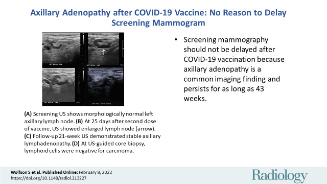
Axillary Adenopathy after COVID-19 Vaccine: No Reason to Delay Screening Mammogram
Affiliations
- PMID: 35133198
- PMCID: PMC8855316
- DOI: 10.1148/radiol.213227
Item in Clipboard
Axillary Adenopathy after COVID-19 Vaccine: No Reason to Delay Screening Mammogram
Radiology.
2022 May.
Erratum in
-
Axillary Adenopathy after COVID-19 Vaccine: No Reason to Delay Screening Mammogram.Radiology. 2022 Sep;304(3):E57. doi: 10.1148/radiol.229015. Radiology. 2022. PMID: 35994402 Free PMC article. No abstract available.
Abstract
Online supplemental material is available for this article.
Conflict of interest statement
Figures
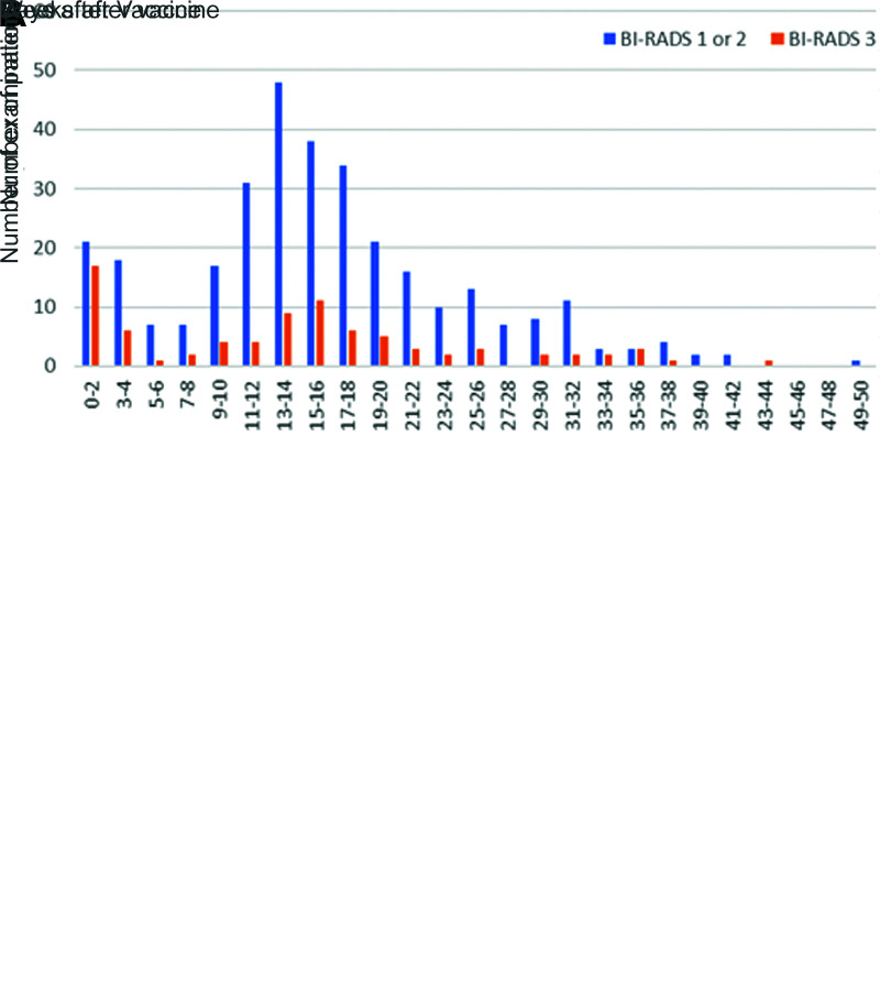
Graphs compare (A) patients with and without lymphadenopathy
(LAD) at initial breast imaging after COVID-19 vaccination. Lymphadenopathy
is seen most commonly in the first 2 weeks after vaccination, but can also
persist at least 10 weeks. (B) Graphs compare patients with LAD
and follow-up imaging. Bars show the percent of examinations assigned Breast
Imaging Reporting and Data System (BI-RADS) category 1 or 2 (negative or
benign findings) versus BI-RADS 3 (probably benign finding; short-term
follow-up is recommended) recommendations by time after the vaccination.
Twenty-five percent of examinations performed at 0–12 weeks were
given BI-RADS 3 recommendations, and none of these patients were
subsequently diagnosed with a new malignancy.
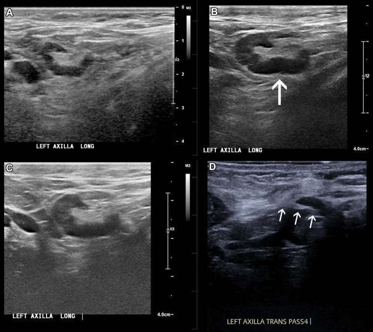
In a 46-year-old patient with a strong family history of breast cancer,
(A) a screening US prior to COVID-19 vaccination
demonstrated a morphologically normal left axillary lymph node.
(B) Twenty-five days following the second dose of the
COVID-19 vaccine, the patient presented with a palpable lump in the left
axilla and US demonstrated enlarged lymph nodes with cortex measuring up to
6 mm in thickness (arrow). (C) Follow-up US 21 weeks following
demonstrated stable axillary lymphadenopathy. (D) A US-guided
core biopsy was then recommended and pathologic analysis demonstrated
lymphoid cells negative for carcinoma. Arrows indicate the path of the
needle.
References
-
- Grimm L , Destounis S , Dogan B , et al. . SBI Recommendations for the Management of Axillary Adenopathy in Patients with Recent COVID-19 Vaccination . Society of Breast Imaging website . https://www.sbi-online.org/Portals/0/Position%20Statements/2021/SBI-reco.... Accessed December 16, 2021 .
MeSH terms
Substances
LinkOut - more resources
Full Text Sources
Medical
