

Effect of a DACC-coated dressing on keratinocytes and fibroblasts in wound healing using an in vitro scratch model
- PMID: 35133505
- PMCID: PMC8825393
- DOI: 10.1007/s10856-022-06648-5
Effect of a DACC-coated dressing on keratinocytes and fibroblasts in wound healing using an in vitro scratch model
Abstract
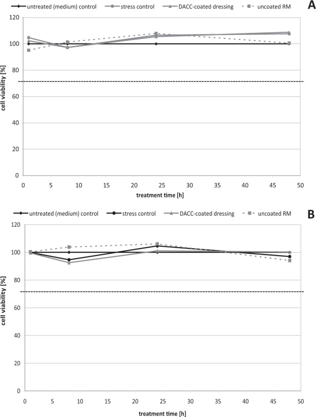
Wound dressings that exert an antimicrobial effect in order to prevent and treat wound infections can be harmful to the wound healing process. Dressings with hydrophobic coatings, however, have been suggested to both reduce the microbial load and promote the healing process. Therefore, the potential effects of a dialkylcarbamoyl chloride (DACC)-coated dressing on fibroblasts and keratinocytes in wound healing were studied using mechanical scratch wounding of confluent cell layers as an in vitro model. Additionally, gene expression analysis by qRT-PCR was used to elucidate the longitudinal effects of the DACC-coated dressing on cell responses, specifically inflammation, growth factor induction and collagen synthesis. DACC promoted cell viability, did not stick to the cell layers, and supported normal wound healing progression in vitro. In contrast, cells became attached to the uncoated reference material, which inhibited scratch closure. Moreover, DACC slightly induced KGF, VEGF, and GM-CSF expression in HaCaT cells and NHDF. Physiological COL1A1 and COL3A1 gene expression by NHDF was observed under DACC treatment with no observable effect on S100A7 and RNASE7 levels in HaCaT cells. Overall, the DACC coating was found to be safe and may positively influence the wound healing outcome. Graphical abstract.
© 2022. The Author(s).
Conflict of interest statement
This study was supported by ABIGO Medical AB Sweden. JH and AA are employees of ABIGO Medical AB.
Figures


Similar articles
-
Significant and rapid reduction of free endotoxin using a dialkylcarbamoyl chloride-coated wound dressing.J Wound Care. 2022 Jun 2;31(6):502-509. doi: 10.12968/jowc.2022.31.6.502. J Wound Care. 2022. PMID: 35678791
-
Antimicrobial effects of bacterial binding to a dialkylcarbamoyl chloride-coated wound dressing: an in vitro study.J Wound Care. 2022 Jul 2;31(7):560-570. doi: 10.12968/jowc.2022.31.7.560. J Wound Care. 2022. PMID: 35797260
-
Effect of a DACC dressing on the growth properties and proliferation rate of cultured fibroblasts.J Wound Care. 2012 Jul;21(7):327-8, 330-2. doi: 10.12968/jowc.2012.21.7.327. J Wound Care. 2012. PMID: 22886332
-
Antimicrobial stewardship strategies in wound care: evidence to support the use of dialkylcarbamoyl chloride (DACC)- coated wound dressings.J Wound Care. 2021 Apr 2;30(4):284-296. doi: 10.12968/jowc.2021.30.4.284. J Wound Care. 2021. PMID: 33856907
-
Bacterial-binding dressings in the management of wound healing and infection prevention: a narrative review.J Wound Care. 2019 Jun 2;28(6):370-382. doi: 10.12968/jowc.2019.28.6.370. J Wound Care. 2019. PMID: 31166862 Review.
Cited by
-
Fabrication and Characterization of Phyllanthus Emblica Extract-Polyvinyl Alcohol/Carboxymethyl Cellulose Sodium Antioxidant Hydrogel and Its Application in Wound Healing.Pharmaceutics. 2024 Nov 29;16(12):1531. doi: 10.3390/pharmaceutics16121531. Pharmaceutics. 2024. PMID: 39771510 Free PMC article.
-
Marine Jellyfish Collagen and Other Bioactive Natural Compounds from the Sea, with Significant Potential for Wound Healing and Repair Materials.Mar Drugs. 2025 Jun 13;23(6):252. doi: 10.3390/md23060252. Mar Drugs. 2025. PMID: 40559661 Free PMC article.
-
Jellyfish Polysaccharides for Wound Healing Applications.Int J Mol Sci. 2022 Sep 29;23(19):11491. doi: 10.3390/ijms231911491. Int J Mol Sci. 2022. PMID: 36232791 Free PMC article.
-
Jellyfishes-Significant Marine Resources with Potential in the Wound-Healing Process: A Review.Mar Drugs. 2023 Mar 24;21(4):201. doi: 10.3390/md21040201. Mar Drugs. 2023. PMID: 37103346 Free PMC article. Review.
References
-
- Christaki E, Marcou M, Tofarides A. Antimicrobial resistance in bacteria: mechanisms, evolution, and persistence. J Mol Evol. 2020;88:26–40. - PubMed
-
- Matzinger P. Tolerance, danger, and the extended family. Annu Rev Immunol. 1994;12:991–1045. - PubMed
-
- Mosti G, Magliaro A, Mattaliano V, Picerni P, Angelotti N. Comparative study of two antimicrobial dressings in infected leg ulcers: a pilot study. J Wound Care. 2015;24:121–7. - PubMed
-
- Doyle RJ. Contribution of the hydrophobic effect to microbial infection. Microbes Infect. 2000;2:391–400. - PubMed
MeSH terms
Substances
LinkOut - more resources
Full Text Sources
Research Materials
Miscellaneous
