

Cerebrospinal fluid kappa free light chains as biomarker in multiple sclerosis-from diagnosis to prediction of disease activity
- PMID: 35133530
- PMCID: PMC9606042
- DOI: 10.1007/s10354-022-00912-7
Cerebrospinal fluid kappa free light chains as biomarker in multiple sclerosis-from diagnosis to prediction of disease activity
Abstract
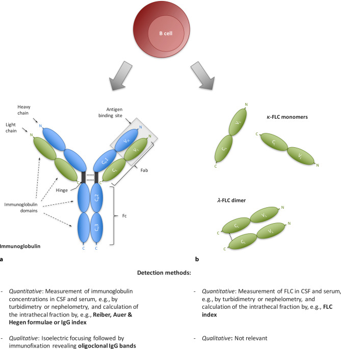
Multiple sclerosis (MS) is a chronic immune-mediated disorder of the central nervous system that shows a high interindividual heterogeneity, which frequently poses challenges regarding diagnosis and prediction of disease activity. In this context, evidence of intrathecal inflammation provides an important information and might be captured by kappa free light chains (κ-FLC) in the cerebrospinal fluid (CSF). In this review, we provide an overview on what is currently known about κ‑FLC, its historical development, the available assays and current evidence on its diagnostic and prognostic value in MS. Briefly, intrathecal κ‑FLC synthesis reaches similar diagnostic accuracy compared to the well-established CSF-restricted oligoclonal bands (OCB) to identify patients with MS, and recent studies even depict its value for prediction of early MS disease activity. Furthermore, detection of κ‑FLC has significant methodological advantages in comparison to OCB detection.
Keywords: Clinically isolated syndrome; FLC index; Oligoclonal bands; Progression; Relapse.
© 2022. The Author(s).
Conflict of interest statement
H. Hegen has participated in meetings sponsored by, received speaker honoraria or travel funding from Bayer, Biogen, Merck, Novartis, Sanofi-Genzyme, Siemens, Teva, and received honoraria for acting as consultant for Biogen, Novartis and Teva. K. Berek has participated in meetings sponsored by and received travel funding from Roche, Teva and Biogen. F. Deisenhammer has participated in meetings sponsored by or received honoraria for acting as an advisor/speaker for Alexion, Almirall, Biogen, Celgene, Genzyme-Sanofi, Merck, Novartis Pharma, Roche, and Teva. His institution has received research grants from Biogen and Genzyme Sanofi. He is section editor of the MSARD Journal (Multiple Sclerosis and Related Disorders).
Figures
References
-
- Compston A, Coles A. Multiple sclerosis. Lancet. 2002;359(9313):1221–1231. - PubMed
-
- Thompson AJ, Banwell BL, Barkhof F, Carroll WM, Coetzee T, Comi G, et al. Diagnosis of multiple sclerosis: 2017 revisions of the McDonald criteria. Lancet Neurol. 2018;17(2):162–173. - PubMed
-
- Arrambide G, Tintore M, Espejo C, Auger C, Castillo M, Río J, et al. The value of oligoclonal bands in the multiple sclerosis diagnostic criteria. Brain. 2018;141(4):1075–1084. - PubMed
-
- Ontaneda D, Tallantyre E, Kalincik T, Planchon SM, Evangelou N. Early highly effective versus escalation treatment approaches in relapsing multiple sclerosis. Lancet Neurol. 2019;18(10):973–980. - PubMed
Publication types
MeSH terms
Substances
LinkOut - more resources
Full Text Sources
Medical
