

Chest CT severity score: assessment of COVID‑19 severity and short-term prognosis in hospitalized Iranian patients
- PMID: 35133531
- PMCID: PMC8824536
- DOI: 10.1007/s10354-022-00914-5
Chest CT severity score: assessment of COVID‑19 severity and short-term prognosis in hospitalized Iranian patients
Abstract
Background: The aim of this study was to evaluate the value of chest computed tomography (CT) severity score in the assessment of coronavirus disease 2019 (COVID‑19) severity and short-term prognosis.
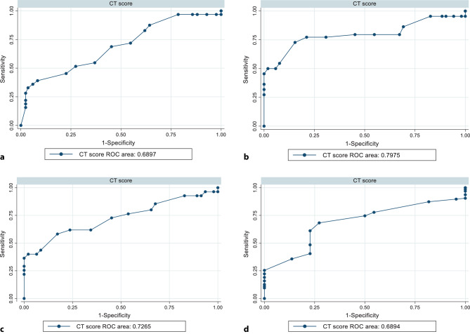
Methods: In this cross-sectional study, we evaluated all patients who were referred to our university hospital, from 21 May 2020 to 22 June 2020 with positive severe acute respiratory syndrome coronavirus 2 (SARS-CoV-2) reverse transcription-polymerase chain reaction (RT-PCR) test. The patients suspected of having other respiratory diseases including influenza, according to an infectious disease specialist, and those without chest CT scan were excluded. A chest CT was obtained for all patients between days 4 and 7 days after symptom onset. Chest CT severity score was also calculated based on the degree of involvement of the lung lobes as 0%, (0 points), 1-25% (1 point), 26-50% (2 points), 51-75% (3 points), and 76-100% (4 points). The CT severity score was quantified by summing the 5 lobe indices (range 0-20). The ROC curve analysis was performed for the clinical value of CT scores in distinguishing the patients based on the severity of disease (mild/moderate group versus severe group), ICU admission, intubation requirement, and mortality.
Results: Of the 148 patients included, 93 patients recovered, while 55 patients died (mortality rate 37%). The area under the curve of CT score for discriminating of recovered patients from deceased individuals was 0.726, and the optimal CT score threshold was 15.5 with 61.8% sensitivity and 76.3% specificity. The best CT score cut-off for discriminating of patients based on the severity of disease was 12.5 with 68.3% sensitivity and 72.7% specificity. In addition, with CT score cut-off of 15.5, sensitivities of 70.8% and 51.6% and specificities of 78% and 72.6% were observed for intubation and ICU admission, respectively.
Conclusion: CT scan and semiquantitative scoring method could be beneficial and applicable in predicting the patient's condition.
Keywords: Computed tomography scan; Computed tomography severity score; Coronavirus disease 2019; Mortality; Severity.
© 2022. The Author(s), under exclusive licence to Springer-Verlag GmbH Austria, ein Teil von Springer Nature.
Conflict of interest statement
The authors declare there they have no conflict of interest.
Figures
Similar articles
-
Predictors of the chest CT score in COVID-19 patients: a cross-sectional study.Virol J. 2021 Nov 18;18(1):225. doi: 10.1186/s12985-021-01699-6. Virol J. 2021. PMID: 34794467 Free PMC article. Review.
-
[Spatial and temporal distribution and predictive value of chest CT scoring in patients with COVID-19].Zhonghua Jie He He Hu Xi Za Zhi. 2021 Mar 12;44(3):230-236. doi: 10.3760/cma.j.cn112147-20200522-00626. Zhonghua Jie He He Hu Xi Za Zhi. 2021. PMID: 33721937 Chinese.
-
Clinical and chest CT features as a predictive tool for COVID-19 clinical progress: introducing a novel semi-quantitative scoring system.Eur Radiol. 2021 Jul;31(7):5178-5188. doi: 10.1007/s00330-020-07623-w. Epub 2021 Jan 15. Eur Radiol. 2021. PMID: 33449185 Free PMC article.
-
Relationship of the cycle threshold values of SARS-CoV-2 polymerase chain reaction and total severity score of computerized tomography in patients with COVID 19.Int J Infect Dis. 2020 Dec;101:160-166. doi: 10.1016/j.ijid.2020.09.1449. Epub 2020 Sep 28. Int J Infect Dis. 2020. PMID: 32992013 Free PMC article.
-
Thoracic imaging tests for the diagnosis of COVID-19.Cochrane Database Syst Rev. 2020 Nov 26;11:CD013639. doi: 10.1002/14651858.CD013639.pub3. Cochrane Database Syst Rev. 2020. Update in: Cochrane Database Syst Rev. 2021 Mar 16;3:CD013639. doi: 10.1002/14651858.CD013639.pub4. PMID: 33242342 Updated.
Cited by
-
Predicting survival of Iranian COVID-19 patients infected by various variants including omicron from CT Scan images and clinical data using deep neural networks.Heliyon. 2023 Nov 8;9(11):e21965. doi: 10.1016/j.heliyon.2023.e21965. eCollection 2023 Nov. Heliyon. 2023. PMID: 38058649 Free PMC article.
-
COVID-19 in Patients with Rheumatic Disease Using Immunomodulatory Drugs: Imaging Findings and Predictors of Hospitalization.Rheumatol Ther. 2022 Dec 2;10(1):249-259. doi: 10.1007/s40744-022-00508-y. eCollection 2023 Feb. Rheumatol Ther. 2022. PMID: 36475037 Free PMC article.
-
Lactate dehydrogenase and PaO2/FiO2 ratio at admission helps to predict CT score in patients with COVID-19: An observational study.J Infect Public Health. 2023 Jan;16(1):136-142. doi: 10.1016/j.jiph.2022.12.009. Epub 2022 Dec 12. J Infect Public Health. 2023. PMID: 36521329 Free PMC article.
-
Evaluation of the value of chest CT severity score in assessment of COVID-19 severity and short-term prognosis.J Family Med Prim Care. 2024 May;13(5):1670-1675. doi: 10.4103/jfmpc.jfmpc_414_23. Epub 2024 May 24. J Family Med Prim Care. 2024. PMID: 38948629 Free PMC article.
-
A Systematic Review of the Relationship between Chest CT Severity Score and Laboratory Findings and Clinical Parameters in COVID-19 Pneumonia.Diagnostics (Basel). 2023 Jun 29;13(13):2223. doi: 10.3390/diagnostics13132223. Diagnostics (Basel). 2023. PMID: 37443616 Free PMC article. Review.
References
MeSH terms
LinkOut - more resources
Full Text Sources
Medical
Miscellaneous
