. 2022 Feb 22;16(2):2928-2941.
doi: 10.1021/acsnano.1c10084. Epub 2022 Feb 8.

Metallointercalators-DNA Tetrahedron Supramolecular Self-Assemblies with Increased Serum Stability

Affiliations

Metallointercalators-DNA Tetrahedron Supramolecular Self-Assemblies with Increased Serum Stability

M Andrey Joaqui-Joaqui et al. ACS Nano. .

Abstract

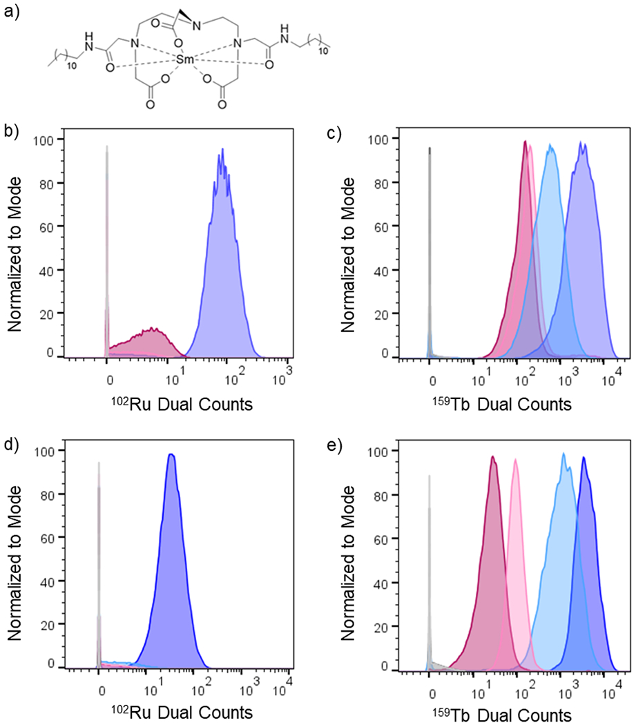
Self-assembly of metallointercalators into DNA nanocages is a rapid and facile approach to synthesize discrete bioinorganic host/guest structures with a high load of metal complexes. Turberfield's DNA tetrahedron can accommodate one intercalator for every two base pairs, which corresponds to 48 metallointercalators per DNA tetrahedron. The affinity of the metallointercalator for the DNA tetrahedron is a function of both the structure of the intercalating ligand and the overall charge of the complex, with a trend in affinity [Ru(bpy)2(dppz)]2+ > [Tb-DOTAm-Phen]3+ ≫ Tb-DOTA-Phen. Intercalation of the metal complex stabilizes the DNA tetrahedron, resulting in an increase of its melting temperature and, importantly, a significant increase in its stability in the presence of serum. [Ru(bpy)2(dppz)]2+, which has a greater affinity for DNA than [Tb-DOTAm-Phen]3+, increases the melting point and decreases degradation in serum to a greater extent than the TbIII complex. In the presence of Lipofectamine, the metallointercalator@DNA nanocage assemblies substantially increase the cell uptake of their respective metal complex. Altogether, the facile incorporation of a large number of metal complexes per assembly, the higher stability in serum, and the increased cell penetration of metallointercalator@DNA make these self-assemblies well-suited as metallodrugs.

Keywords: DNA tetrahedron; cell uptake; metallointercalator; serum stability; supramolecular self-assembly.


Conflict of interest statement

The authors declare no competing financial interest.

Figures

Figure 1.
Figure 1.
Chemical structures of [Ru(bpy)2(dppz)]2+, [Tb-DOTAm-Phen]3+ and Tb-DOTA-Phen.
Figure 2.
Figure 2.
Supramolecular functionalization of DNA tetrahedron structures with metallointercalators.
Figure 3.
Figure 3.
(a) Fluorescence spectra of [Ru(bpy)2(dppz)]2+ upon addition of increasing concentrations of DNA tetrahedron. (b) Time-delayed phosphorescence spectra of [Tb-DOTAm-Phen]3+ upon addition of increasing concentrations of DNA tetrahedron. (c) Normalized luminescence intensity as a function of the ratio of DNA base pair (b.p.) to metallointercalator (ML). Error bars represent ±1 standard deviation (n = 3). I = integrated luminescence intensity of the metallointercalator upon addition of increasing amounts of DNA tetrahedron, I0 = integrated luminescence intensity of the metallointercalator in the absence of DNA tetrahedron. Experimental conditions: [metallointercalator] = 10 μM in buffer ([Tris] = 10 mM, [MgCl2] = 5 mM, pH 7.4); T = 20 °C. For [Ru(bpy)2(dppz)]2+: λex = 440 nm, fluorescence is integrated from λem= 500 nm to 800 nm, excitation slit width = 10 nm and emission slit width = 10 nm. For [Tb-DOTAm-Phen]3+: λex = 345 nm, time-delayed luminesce is integrated from λem= 450 nm to 650 nm, delay time = 0.1 ms, gate time = 5 ms, excitation slit width = 5 nm and emission slit width = 5 nm.
Figure 4.
Figure 4.
Job’s plot of the DNA tetrahedron responsive [Ru(bpy)2(dppz)]2+. I=integrated luminescence intensity from 500 nm to 800 nm. I0 = integrated luminescence intensity in the absence of the DNA tetrahedron from 500 nm to 800 nm. Experimental conditions: Total concentration of [Ru(bpy)2(dppz)]2+ + DNA base pair (b.p) = 10 μM in buffer ([Tris] = 10 mM, [MgCl2] = 5 mM, pH 7.4); T = 20 °C; λex = 440 nm, excitation slit width = 10 nm; emission slit width = 10 nm.
Figure 5.
Figure 5.
Displacement of [Ru(bpy)2(dppz)]2+ from the DNA tetrahedron upon addition of competing terbium-based metallointercalators. [Tb-L]= concentration of corresponding [Tb-DOTAm-Phen]3+ and [Tb-DOTA-Phen]. I = integrated luminescence intensity of [Ru(bpy)2(dppz)]+2@DNA upon addition of increasing amounts of competing metallointercalator, I0 = integrated luminescence intensity of [Ru(bpy)2(dppz)]+2@DNA tetrahedron in absence of competing metallointercalator. Error bars represent ±1 standard deviation (n = 3). Experimental conditions: [Ru(bpy)2(dppz)]+2 (10 μM) and DNA tetrahedron (20 μM b.p) in M in buffer ([Tris] = 10 mM, [MgCl2] = 5 mM, pH 7.4); T = 20 °C. Fluorescence integrated from λem = 500 nm to 800 nm, λex = 440 nm, excitation slit width = 10 nm; emission slit width = 10 nm.
Figure 6.
Figure 6.
Representative melting curves of DNA tetrahedron and metallointercalator@DNA tetrahedron as measured by UV-spectroscopy and fluorescence. In all cases melting point was calculated as the average of three different measurements. Experimental conditions for UV-melting curves: Absorbance recorded at 260 nm. [DNA tetrahedron] = 150 nM, [metallointercalator] = 7.7 μM in buffer ([Tris] = 10 mM, [MgCl2] = 5 mM, pH 7.4), Heating rate = 1 °C/min. Experimental conditions for fluorescence-melting curve: λem = 618 nm, λex = 440 nm, I= luminescence intensity of [Ru(bpy)2(dppz)]2+@DNA, I0 = luminescence intensity of [Ru(bpy)2(dppz)]2+ in absence of DNA at the same temperature. [DNA tetrahedron] = 30 nM, [ [Ru(bpy)2(dppz)]2+ ] = 1.5 μM in buffer ([Tris] = 10 mM, [MgCl2] = 5 mM, pH 7.4), excitation slit width = 10 nm, emission slit width = 10 nm. Heating rate = 1 °C/min.
Figure 7.
Figure 7.
Atomic force microscopy images of DNA tetrahedron with or without metal complex. a) DNA tetrahedron, b) [Ru(bpy)2(dppz)]2+@DNA assembly, c) [Tb-DOTAm-Phen]3+@DNA assembly.
Figure 8.
Figure 8.
Stability of DNA tetrahedron and metallointercalator@DNA tetrahedron assemblies in: a) 10% fetal bovine serum (FBS), b) 10% mouse serum. Percentage of intact DNA tetrahedron was estimated by non-denaturing agarose gel electrophoresis 2% in TBE 1X, visualized with SYBR Safe staining, and plotted as a function of time. Error bars represent ±1 standard deviation (n = 3).
Figure 9.
Figure 9.
a) Chemical structure Sm-DDD probe for cell labeling by cytometry analysis. b) Histogram showing 102Ru dual count intensity distribution for L6 cells treated for 4 hours with [Ru(bpy)2(dppz)]2+, Lipofectamine + [Ru(bpy)2(dppz)]2+, [Ru(bpy)2(dppz)]2+@DNA, Lipofectamine + [Ru(bpy)2(dppz)]2+@DNA, and controls. c) Histogram showing 159Tb dual count intensity distribution for L6 cells treated for 4 hours with [Tb-DOTAm-Phen]3+, Lipofectamine + [Tb-DOTAm-Phen]3+, [Tb-DOTAm-Phen]3+@DNA, Lipofectamine + [Tb-DOTAm-Phen]3+@DNA and controls. d) Histogram showing 102Ru dual count intensity distribution for HEK-293 cells treated for 4 hours with [Ru(bpy)2(dppz)]2+, Lipofectamine + [Ru(bpy)2(dppz)]2+, [Ru(bpy)2(dppz)]2+@DNA, Lipofectamine + [Ru(bpy)2(dppz)]2+@DNA, and controls. c) Histogram showing 159Tb dual count intensity distribution for HEK-293 cells treated for 4 hours with [Tb-DOTAm-Phen]3+, Lipofectamine + [Tb-DOTAm-Phen]3+, [Tb-DOTAm-Phen]3+@DNA, Lipofectamine + [Tb-DOTAm-Phen]3+@DNA and controls. Legend for samples colors is indicated in table 2.

Similar articles

Cited by

References

    1. Simoncelli S; Li Y; Cortés E; Maier SA Nanoscale Control of Molecular Self-Assembly Induced by Plasmonic Hot-Electron Dynamics. ACS Nano. 2018, 12, 2184–2192. - PubMed
    1. Whitesides GM; Mathias JP; Seto CT Molecular Self-Assembly and Nanochemistry: A Chemical Strategy for the Synthesis of Nanostructures. Science. 1991, 254, 1312–1319. - PubMed
    1. Ariga K; Nishikawa M; Mori T; Takeya J; Shrestha LK; Hill JP Self-Assembly As a Key Player for Materials Nanoarchitectonics. Sci. Technol. Adv. Mater 2019, 20, 51–95. - PMC - PubMed
    1. Zhang S Fabrication of Novel Biomaterials Through Molecular Self-Assembly. Nat. Biotechnol 2003, 21, 1171–1178. - PubMed
    1. Tabacchi G Supramolecular Organization in Confined Nanospaces. ChemPhysChem. 2018, 19, 1249–1297. - PubMed

Publication types