Review
. 2022 Sep 14;122(17):14055-14065.
doi: 10.1021/acs.chemrev.1c00753. Epub 2022 Feb 8.

Cryo-EM of Helical Polymers

Affiliations
Review

Cryo-EM of Helical Polymers

Fengbin Wang et al. Chem Rev. .

Abstract

While the application of cryogenic electron microscopy (cryo-EM) to helical polymers in biology has a long history, due to the huge number of helical macromolecular assemblies in viruses, bacteria, archaea, and eukaryotes, the use of cryo-EM to study synthetic soft matter noncovalent polymers has been much more limited. This has mainly been due to the lack of familiarity with cryo-EM in the materials science and chemistry communities, in contrast to the fact that cryo-EM was developed as a biological technique. Nevertheless, the relatively few structures of self-assembled peptide nanotubes and ribbons solved at near-atomic resolution by cryo-EM have demonstrated that cryo-EM should be the method of choice for a structural analysis of synthetic helical filaments. In addition, cryo-EM has also demonstrated that the self-assembly of soft matter polymers has enormous potential for polymorphism, something that may be obscured by techniques such as scattering and spectroscopy. These cryo-EM structures have revealed how far we currently are from being able to predict the structure of these polymers due to their chaotic self-assembly behavior.


Figures

Figure 1)
Figure 1)
Helical symmetry is best understood in terms of the helical net. (a) An illustration of a helix without point group symmetry (rise: 4 Å, twist: 55°, C1). The asymmetric units (ASU) are represented by spheres. The right-handed 1-start helix that passes through every subunit is shown as a dashed line. (b) The helical net of a, using the convention that the surface is unrolled and we are viewing it from the outside. The helix crosses the horizontal red line once, so the helix is called a 1-start. The helical rise and helical twist along the right-handed 1-start helix are labeled. (c) An illustration of a helix with point group symmetry (rise: 4 Å, twist: 55°, C5). Because of the rotational point group symmetry, any rotation of this structure by multiples of 72° is an identity operation. The asymmetric units are represented by spheres. The subunits along a single 5-start strand are shown in red, and the dashed lines show the 5-start helices. (d) The helical net of c, using the convention above. The right-handed 5-start helices cross the horizontal red line five times, hence the name 5-start.
Figure 2)
Figure 2)
Cryo-EM has become the method of choice for helical polymers. Cryo-EM maps better than 4 Å resolution, now routinely attainable, clearly resolve some side chains of the peptide or peptide-like compounds, and one has the ability to build atomic models de novo. (a) Cryo-EM reconstruction of a cross-α nanotube at 3.8 Å resolution. The α-helices form stacks, and the three stacks forming the tube are each shown in a different color. The ASU is a single α-helical peptide containing 36 residues. The density from a single subunit is shown on the right as a grey mesh, and a ribbon diagram of the atomic model has been fit into this density. (b) Cryo-EM reconstruction of a cross-β nanotube at 3.5 Å resolution. The β-sheets on the inside (yellow) are parallel, and those on the outside (cyan) are antiparallel. The ASU contains four peptides, shown on the right, forming four β-strands. Although each 8-residue peptide is chemically identical, each of the four peptides in the ASU is in a different environment.
Figure 3)
Figure 3)
The loss of information about the absolute hand in projection images is best illustrated with a clinical x-ray of the human hand, since such x-ray images, like cryo-EM images, are projections. From a radiograph on the left, one cannot tell whether this is a right hand, palm down, or a left hand, palm up. The image on the right is simply the mirror image of the one on the left, and this could be either a left hand, palm down, or a right hand, palm up. Radiograph courtesy of Department of Radiology, University of California San Diego.
Figure 4)
Figure 4)
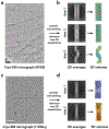
Polymorphism is the norm rather than the exception with many peptide assemblies, and cryo-EM can sort out heterogeneity of polymers. (a) & (c), representative cryo-EMs of KFE8 nanotube (a) and 1-KMe3 nanotube (c). Scale bar is 50 nm. (b) & (d), After automatic particle picking and reference-free 2D classfications, helical polymers are grouped into different classes. The differences are obvious by looking at the averages, and helical symmetry determination and high-resolution reconstructions can subsequently be done for each class. In (b) class 1 corresponds to tubes with five sheets, class 2 corresponds to tubes formed from four sheets, and class 3 is a ribbon containing a single sheet. In (d), class 1 corresponds to a filament with seven peptides per plane, and class 2 corresponds to a filament with six peptides per plane.
Figure 5)
Figure 5)
Helical polymers made from small subunits can easily adopt different helical symmetries while maintaining relatively conserved interactions. Two peptide examples are shown here: (a)-(d) nanotube 29–20-2 compared with AS2; (e)-(h) nanotube Form I compared with F1-N2. (a) & (e), The amino acid sequences of the peptides. The dots between the sequences indicate the residues that are non-identical in each of the pairs. (b) & (f), The determined helical symmetry and atomic models of the nanotubes (PDB 7RX4 for AS2 and 7RX5 for F1-N2). (c) & (g), The interface comparison between two nanotubes shows that relatively conserved contacts are maintained, even though the helical symmetry has changed. (d) & (h), The averaged power spectrum (only showing one of two, as they look similar) of the nanotubes, and the helical indexing.
Figure 6)
Figure 6)
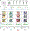
Ambiguities frequently exist in helical symmetry determination. (a), The averaged power spectrum of nanotube F1-N2, and four possible helical symmetries generated from indexing the power spectrum. (b), The “gold-standard” FSCs after applying those four different symmetries in helical reconstructions are nearly indistinguishable, despite the fact that three of these symmetries are wrong. (c)-(e), The reconstruction volumes for those four different symmetries filtered to resolutions of 8 Å (c), 5 Å (d), and 3.5 Å (e). At 5 Å resolution or worse all four symmetries generate maps that might be considered reasonable for α-helical subunits. (f), The density of a single helix is shown from the 3.5 Å map. The comparison between the density and the atomic model shows that symmetry 3 is the correct one. (g), The model:map FSC for those four maps with different symmetries and corresponding atomic models. The commonly used arbitrary cutoffs (0.5 and 0.38) are shown. Using the FSC=0.38 criterion (where 0.38 =√ 0.143), only one of these (symmetry 1) might be excluded as obviously incorrect.

Similar articles

Cited by

References

    1. Kuhlbrandt W Biochemistry. The resolution revolution. Science 2014, 343, 1443–1444. - PubMed
    1. Bai XC; McMullan G; Scheres SH How cryo-EM is revolutionizing structural biology. Trends Biochem. Sci. 2015, 40, 49–57. - PubMed
    1. Egelman EH The Current Revolution in Cryo-EM. Biophys. J. 2016, 110, 1008–1012. - PMC - PubMed
    1. Busiek KK; Margolin W Bacterial actin and tubulin homologs in cell growth and division. Curr. Biol. 2015, 25, R243–R254. - PMC - PubMed
    1. Egelman EH Cryo-EM of bacterial pili and archaeal flagellar filaments. Curr. Opin. Struct. Biol. 2017, 46, 31–37. - PMC - PubMed

Publication types