Review
. 2022 Mar 8;7(5):e157342.
doi: 10.1172/jci.insight.157342.

Disulfiram inhibits neutrophil extracellular trap formation and protects rodents from acute lung injury and SARS-CoV-2 infection

Affiliations
Review

Disulfiram inhibits neutrophil extracellular trap formation and protects rodents from acute lung injury and SARS-CoV-2 infection

Jose M Adrover et al. JCI Insight. .

Abstract

Severe acute lung injury has few treatment options and a high mortality rate. Upon injury, neutrophils infiltrate the lungs and form neutrophil extracellular traps (NETs), damaging the lungs and driving an exacerbated immune response. Unfortunately, no drug preventing NET formation has completed clinical development. Here, we report that disulfiram - an FDA-approved drug for alcohol use disorder - dramatically reduced NETs, increased survival, improved blood oxygenation, and reduced lung edema in a transfusion-related acute lung injury (TRALI) mouse model. We then tested whether disulfiram could confer protection in the context of SARS-CoV-2 infection, as NETs are elevated in patients with severe COVID-19. In SARS-CoV-2-infected golden hamsters, disulfiram reduced NETs and perivascular fibrosis in the lungs, and it downregulated innate immune and complement/coagulation pathways, suggesting that it could be beneficial for patients with COVID-19. In conclusion, an existing FDA-approved drug can block NET formation and improve disease course in 2 rodent models of lung injury for which treatment options are limited.

Keywords: COVID-19; Immunology; Innate immunity; Neutrophils.


Conflict of interest statement

Conflict of interest: ME is a member of the research advisory board for brensocatib for Insmed Inc., a member of the scientific advisory board for Vividion Therapeutics Inc., and a consultant for Protalix Inc. RES is on the scientific advisory board of Miromatrix Inc. and is a consultant and speaker for Alnylam Inc.

Figures

Figure 1
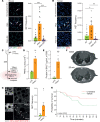
Figure 1. Disulfiram blocks neutrophil extracellular trap (NET) formation, and TRALI is a model of NET-driven lung injury.
(A) Ex vivo NET formation assay of mouse neutrophils sorted by FACS, unstimulated/untreated (NT) or stimulated with 100 nM of PMA or PMA + 10 μM disulfiram (PMA+DS). NET frequency (NET counts normalized to neutrophil counts, with NETs defined by the triple colocalization events of DNA, myeloperoxidase [MPO], and citrullinated histone H3 [citH3]). n = 18 random fields from 4 mice per condition. (B) Ex vivo NET formation assay of human neutrophils from RBC-lysed blood, unstimulated or stimulated with PMA or PMA + 10 μM disulfiram (PMA+DS). n = 18 random fields from 3 healthy donors per condition. Scale bar: 50 μm. (C) Experimental design used to induce TRALI. (D) Absolute number of neutrophils (PMNs) infiltrated to the lung upon TRALI, determined by flow cytometry. n = 4 mice per group. (E) Protein content in the bronchoalveolar lavage fluid (BALF) as a measure of endothelial integrity. n = 6 control and 8 TRALI mice. (F) Representative longitudinal CT scan of a mouse subjected to TRALI showing edema formation over time (representative of CT scans from 11 independent mice). (G) Whole mount tissue clearing images (left, showing CD31 and NETs, defined as the triple colocalization channel of DNA, MPO, and citH3). Quantification (right) of NETs in the lungs of mice 40 minutes after TRALI induction or in mice treated only with LPS. n = 6 lungs per group. Scale bar: 100 μm. (H) Survival of mice after TRALI induction and treatment with Cl-amidine, a PAD4 inhibitor able to block NET formation, or vehicle. n = 27 (vehicle) and 24 (Cl-amidine) mice. Data are shown as mean ± SEM. *P < 0.05, **P < 0.01, ***P < 0.001; as determined by unpaired 2-tailed t test analysis (D, E, and G), 1-way ANOVA with Tukey’s multiple comparison test (A and B), or log-rank (Mantel-Cox) test (H). Arrows indicate NETs in A, B, and G.
Figure 2
Figure 2. Disulfiram blocks NET formation in vivo and protects against acute lung injury.
(A) Experimental design. (B) Whole mount tissue clearing images (left) and quantification (right) of NETs formed in vivo upon TRALI induction in mice treated with disulfiram or vehicle. n = 10 lung volumes from 7 mice per group. Scale bar: 50 μm. Arrows point to NETs. (C) Survival curve of mice treated with 50 mg/kg disulfiram in sesame oil 24 hours and 3 hours before TRALI induction. n = 20 mice per group. (D) IL-1β measurement in lung lysates of LPS-only–treated control mice or mice subject to TRALI induction and treated with vehicle or disulfiram. n = 5 mice per group, lungs acquired 40 minutes after TRALI induction. (E) Survival curves of mice treated i.v. with 50 μg of IL-1β blocking antibodies or isotype control antibodies 5 minutes prior to TRALI induction. n = 20 mice per group. Data are shown as mean ± SEM. **P < 0.01, as determined by 1-way ANOVA with Tukey’s multiple comparison test (D) or unpaired 2-tailed Student’s t test (B). Survival plots show the probability of survival as determined by log-rank (Mantel-Cox) test (C and E).
Figure 3
Figure 3. Disulfiram treatment improves key respiratory parameters upon TRALI induction.
(A) pO2 measured longitudinally on surviving mice after TRALI induction and treatment with disulfiram or vehicle. n = 4 (vehicle) and 3 (disulfiram) mice. (B) Protein content in the BALF of naive mice or mice after TRALI induction and treatment with either disulfiram or vehicle. n = 5 mice per condition. (C) Representative projections from longitudinal CT scans of mice after TRALI induction and treatment with disulfiram or vehicle, showing the lung volume (in blue) and water-dense tissue (edema, in red). Representative of n = 10 mice per group. (D) Quantification of the longitudinal CT scans of mice after TRALI induction and treatment with disulfiram or vehicle. Basal HU units (prior to TRALI induction) were subtracted from all subsequent measurements to represent the increase in edema formation. n = 10 mice per group. Data are shown as mean ± SEM. *P < 0.05, ***P < 0.001, as determined by 1-way ANOVA with Tukey’s multiple comparison test (B) or 2-way ANOVA (A and D).
Figure 4
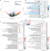
Figure 4. RNA-Seq data from lungs of infected hamsters treated with disulfiram or vehicle.
(A) Volcano plot of log2FC (log2 of the fold change) versus –log10P (log10 of the P value) of all genes in the data set. Positive and negative values on the x axis represent genes upregulated and downregulated, respectively, by disulfiram treatment. Green dots show genes with a log2 fold change over 2 between conditions, blue dots represent genes with a P value under 0.05, and red dots show those genes that have both a log2 fold change > 2 and P < 0.05. (B) GO biological processes terms enriched in the whole differentially expressed genes list highlighting some of the terms (full list in Supplemental Table 2) related to immune functions (blue), response to oxygen levels (orange), other (black), and viral life cycle (red). (C) Clustering of Reactome pathways enriched in the genes downregulated (left) or upregulated (right) in response to disulfiram in SARS-CoV-2–infected golden hamsters. Some of the clusters (gray squares) are shown here (full list in Supplemental Table 4). Color and bubble size reflect the –log10 of the P value for that pathway and the number of genes present in the data set belonging to a particular pathway, respectively.
Figure 5
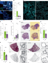
Figure 5. Disulfiram improves lung histology in a golden hamster SARS-CoV-2 infection model.
(A) Representative images from whole mount cleared SARS-CoV-2–infected lungs from hamsters treated with disulfiram or vehicle. Arrows point to NETs, defined as triple colocalization events of DNA, MPO, and citH3. Representative of 5 independent whole mounts per group. (B) Quantification of NETs in the lungs of SARS-CoV-2–infected hamsters. A group was started on daily disulfiram treatment 24 hours prior to infection (pretreat.), while disulfiram was initiated in the other group one day post infection (post.). n = 10 lung volumes from 5 hamsters per group. (C and D) Representative images (showing MPO in cyan) and quantification of neutrophil infiltration to the lungs of SARS-CoV-2–infected hamsters. n = 30 random fields from 5 lungs per group. (E) Quantification of SARS-CoV-2 nucleocapsid signal normalized to β-actin (both proteins detected in lung lysates by Western blot) in disulfiram- and vehicle-treated hamsters, showing that disulfiram does not affect viral load. n = 5 Western blots from 3 uninfected hamsters, 5 Western blots from 5 infected and vehicle-treated hamsters, and 10 Western blots from infected and disulfiram-treated hamsters (5 from the pretreatment and 5 from the posttreatment groups). (F and G) Representative images (left, original image; right, detection overlay showing infiltrated area in violet) and quantification of the heavily immune-infiltrated areas from H&E-stained lungs of disulfiram- or vehicle-treated hamsters infected with SARS-CoV-2. n = 5 (vehicle) and 10 (disulfiram) lungs per group. (H and I) Quantification (mean value of 10 independent measurements per lung) and representative images of perivascular fibrosis in the Masson trichrome–stained lungs of infected hamsters treated with disulfiram or vehicle. n = 5 (vehicle) and 10 (disulfiram) lungs per group. Data are shown as mean ± SEM. *P < 0.05, ***P < 0.001 as determined by 1-way ANOVA with Tukey’s multiple comparison test (A) or unpaired 2-tailed t test analysis (D, E, G, and H). Scale bars: (A) 25 μm, (C) 50 μm, (F and I) 1 mm.

References

    1. Rubenfeld GD, et al. Incidence and outcomes of acute lung injury. N Engl J Med. 2005;353(16):1685–1693. doi: 10.1056/NEJMoa050333. - DOI - PubMed
    1. Cheng OZ, Palaniyar N. NET balancing: a problem in inflammatory lung diseases. Front Immunol. 2013;4:1. - PMC - PubMed
    1. Adrover JM, et al. Programmed ‘disarming’ of the neutrophil proteome reduces the magnitude of inflammation. Nat Immunol. 2020;21(2):135–144. doi: 10.1038/s41590-019-0571-2. - DOI - PMC - PubMed
    1. Thomas GM, et al. Extracellular DNA traps are associated with the pathogenesis of TRALI in humans and mice. Blood. 2012;119(26):6335–6343. doi: 10.1182/blood-2012-01-405183. - DOI - PMC - PubMed
    1. Branzk N, et al. Neutrophils sense microbial size and selectively release neutrophil extracellular traps in response to large pathogens. Nat Immunol. 2015;15(11):1017–1025. - PMC - PubMed

Publication types

Substances