T cell response to intact SARS-CoV-2 includes coronavirus cross-reactive and variant-specific components
- PMID: 35133988
- PMCID: PMC8986086
- DOI: 10.1172/jci.insight.158126
T cell response to intact SARS-CoV-2 includes coronavirus cross-reactive and variant-specific components
Abstract
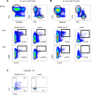
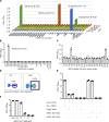
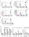
SARS-CoV-2 provokes a robust T cell response. Peptide-based studies exclude antigen processing and presentation biology, which may influence T cell detection studies. To focus on responses to whole virus and complex antigens, we used intact SARS-CoV-2 and full-length proteins with DCs to activate CD8 and CD4 T cells from convalescent people. T cell receptor (TCR) sequencing showed partial repertoire preservation after expansion. Resultant CD8 T cells recognize SARS-CoV-2-infected respiratory tract cells, and CD4 T cells detect inactivated whole viral antigen. Specificity scans with proteome-covering protein/peptide arrays show that CD8 T cells are oligospecific per subject and that CD4 T cell breadth is higher. Some CD4 T cell lines enriched using SARS-CoV-2 cross-recognize whole seasonal coronavirus (sCoV) antigens, with protein, peptide, and HLA restriction validation. Conversely, recognition of some epitopes is eliminated for SARS-CoV-2 variants, including spike (S) epitopes in the Alpha, Beta, Gamma, and Delta variant lineages.
Keywords: Antigen-presenting cells; Dendritic cells; Infectious disease; T cells.
Figures


Update of
-
T cell response to intact SARS-CoV-2 includes coronavirus cross-reactive and variant-specific components.medRxiv [Preprint]. 2022 Jan 25:2022.01.23.22269497. doi: 10.1101/2022.01.23.22269497. medRxiv. 2022. Update in: JCI Insight. 2022 Mar 22;7(6):e158126. doi: 10.1172/jci.insight.158126. PMID: 35118477 Free PMC article. Updated. Preprint.
Similar articles
-
T cell response to intact SARS-CoV-2 includes coronavirus cross-reactive and variant-specific components.medRxiv [Preprint]. 2022 Jan 25:2022.01.23.22269497. doi: 10.1101/2022.01.23.22269497. medRxiv. 2022. Update in: JCI Insight. 2022 Mar 22;7(6):e158126. doi: 10.1172/jci.insight.158126. PMID: 35118477 Free PMC article. Updated. Preprint.
-
Breadth and Durability of SARS-CoV-2-Specific T Cell Responses following Long-Term Recovery from COVID-19.Microbiol Spectr. 2023 Aug 17;11(4):e0214323. doi: 10.1128/spectrum.02143-23. Epub 2023 Jul 10. Microbiol Spectr. 2023. PMID: 37428088 Free PMC article.
-
Immunopeptidome profiling of human coronavirus OC43-infected cells identifies CD4 T cell epitopes specific to seasonal coronaviruses or cross-reactive with SARS-CoV-2.bioRxiv [Preprint]. 2022 Dec 1:2022.12.01.518643. doi: 10.1101/2022.12.01.518643. bioRxiv. 2022. Update in: PLoS Pathog. 2023 Jul 27;19(7):e1011032. doi: 10.1371/journal.ppat.1011032. PMID: 36482973 Free PMC article. Updated. Preprint.
-
SARS-CoV-2-Seronegative Subjects Target CTL Epitopes in the SARS-CoV-2 Nucleoprotein Cross-Reactive to Common Cold Coronaviruses.Front Immunol. 2021 Apr 28;12:627568. doi: 10.3389/fimmu.2021.627568. eCollection 2021. Front Immunol. 2021. PMID: 33995351 Free PMC article. Clinical Trial.
-
A systemic review of T-cell epitopes defined from the proteome of SARS-CoV-2.Virus Res. 2023 Jan 15;324:199024. doi: 10.1016/j.virusres.2022.199024. Epub 2022 Dec 13. Virus Res. 2023. PMID: 36526016 Free PMC article.
Cited by
-
Viral Shedding 1 Year Following First-Episode Genital HSV-1 Infection.JAMA. 2022 Nov 1;328(17):1730-1739. doi: 10.1001/jama.2022.19061. JAMA. 2022. PMID: 36272098 Free PMC article.
-
The Influence of Cross-Reactive T Cells in COVID-19.Biomedicines. 2024 Mar 2;12(3):564. doi: 10.3390/biomedicines12030564. Biomedicines. 2024. PMID: 38540178 Free PMC article. Review.
-
Short Peptides as Powerful Arsenal for Smart Fighting Cancer.Cancers (Basel). 2024 Sep 24;16(19):3254. doi: 10.3390/cancers16193254. Cancers (Basel). 2024. PMID: 39409876 Free PMC article. Review.
-
The prospect of universal coronavirus immunity: characterization of reciprocal and non-reciprocal T cell responses against SARS-CoV2 and common human coronaviruses.Front Immunol. 2023 Oct 13;14:1212203. doi: 10.3389/fimmu.2023.1212203. eCollection 2023. Front Immunol. 2023. PMID: 37901229 Free PMC article.
-
Evidence for broad cross-reactivity of the SARS-CoV-2 NSP12-directed CD4+ T-cell response with pre-primed responses directed against common cold coronaviruses.Front Immunol. 2023 May 5;14:1182504. doi: 10.3389/fimmu.2023.1182504. eCollection 2023. Front Immunol. 2023. PMID: 37215095 Free PMC article.
References
-
- Snyder TM, et al. Magnitude and dynamics of the T-cell response to sars-cov-2 infection at both individual and population levels [preprint]. Posted on medRxiv September 17, 2020. - DOI
Publication types
Grants and funding
LinkOut - more resources
Full Text Sources
Research Materials
Miscellaneous

