. 2022 Feb 8;18(2):e1010248.
doi: 10.1371/journal.ppat.1010248. eCollection 2022 Feb.

A SARS-CoV-2 variant elicits an antibody response with a shifted immunodominance hierarchy

Affiliations

A SARS-CoV-2 variant elicits an antibody response with a shifted immunodominance hierarchy

Allison J Greaney et al. PLoS Pathog. .

Abstract

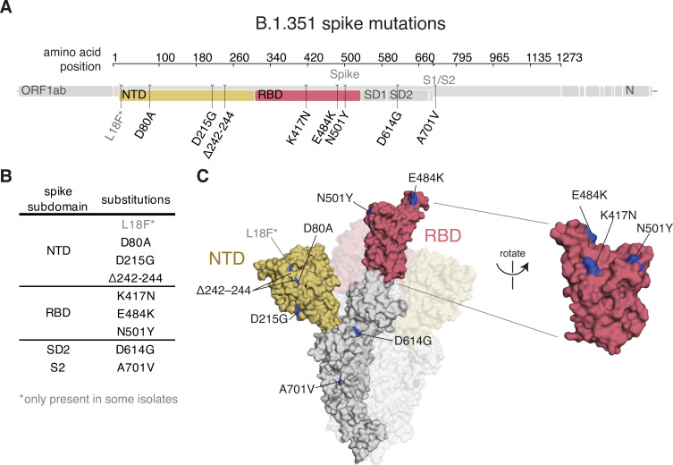
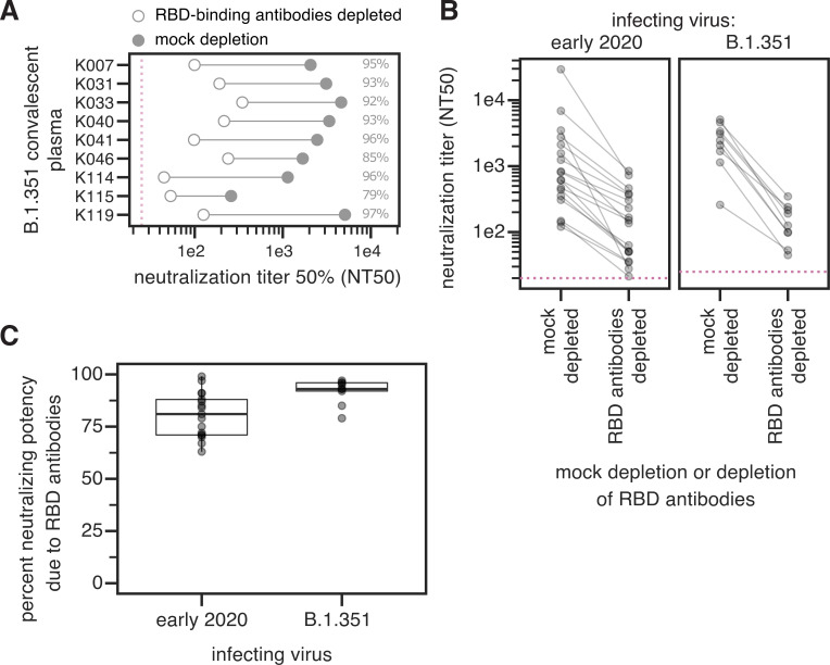
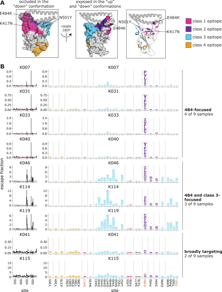
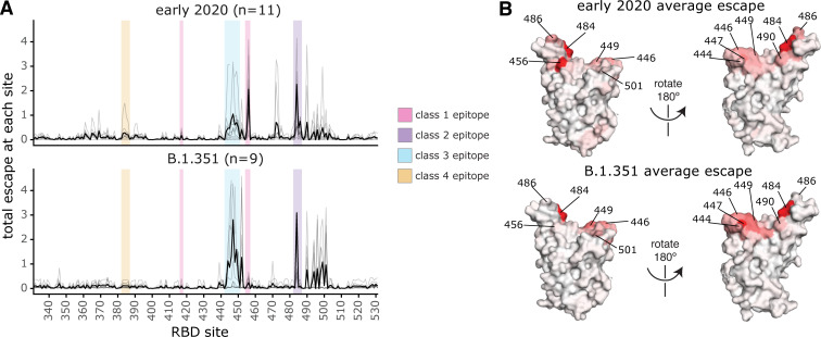
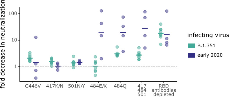
Many SARS-CoV-2 variants have mutations at key sites targeted by antibodies. However, it is unknown if antibodies elicited by infection with these variants target the same or different regions of the viral spike as antibodies elicited by earlier viral isolates. Here we compare the specificities of polyclonal antibodies produced by humans infected with early 2020 isolates versus the B.1.351 variant of concern (also known as Beta or 20H/501Y.V2), which contains mutations in multiple key spike epitopes. The serum neutralizing activity of antibodies elicited by infection with both early 2020 viruses and B.1.351 is heavily focused on the spike receptor-binding domain (RBD). However, within the RBD, B.1.351-elicited antibodies are more focused on the "class 3" epitope spanning sites 443 to 452, and neutralization by these antibodies is notably less affected by mutations at residue 484. Our results show that SARS-CoV-2 variants can elicit polyclonal antibodies with different immunodominance hierarchies.


Conflict of interest statement

I have read the journal’s policy and the authors of this manuscript have the following competing interests: J.D.B. consults for Moderna and Flagship Labs 77 on topics related to viral evolution, and has the potential to receive a share of IP revenue as an inventor on a Fred Hutch optioned technology/patent (application WO2020006494) related to deep mutational scanning of viral proteins. A.J.G, T.N.S., and J.D.B have the potential to receive a share of IP revenue as an inventor on a Fred Hutch optioned technology related to deep mutational scanning of the receptor-binding domain of SARS-CoV-2 Spike protein. H.Y.C. is a consultant for Merck, Pfizer, Ellume, and the Bill and Melinda Gates Foundation and has received support from Cepheid and Sanofi-Pasteur. D.V. is named as an inventor on a patent application filed by the University of Washington related to SARS-CoV-2 vaccines and has received an unrelated sponsored research agreement from Vir Biotechnology Inc. D.C. is an employee of and may hold shares in Vir Biotechnology. The other authors declare no competing interests.

Figures

Fig 1
Fig 1. B.1.351 spike mutations.
(A, B) Mutations in the B.1.351 spike relative to Wuhan-Hu-1 [17]. L18F is only present in some B.1.351 isolates. Visualization generated by https://covdb.stanford.edu/sierra/sars2/by-patterns/. (C) Sites where mutations occur in the spike ectodomain are highlighted in blue on the Wuhan-Hu-1 one-RBD open spike trimer (left, PDB 6ZGG) [75] or RBD (right) (PDB 6M0J) [76]. The surface of one spike monomer is shown; the other two protomers are transparent.
Fig 2
Fig 2. The neutralizing activity of plasma antibodies elicited by B.1.351 infection is heavily focused on the RBD.
(A) The neutralizing titer (NT50) of plasmas from B.1.351-infected individuals against B.1.351 spike-pseudotyped lentiviral particles, following mock depletion or depletion of B.1.351 RBD-binding antibodies. (B) Comparison of neutralization titer following mock depletion or depletion of B.1.351 RBD-binding antibodies for early 2020 (n = 17) [28] and B.1.351 convalescent plasmas (n = 9). The pink dashed line in A, B indicates the limit of detection (NT50 of 25 for B.1.351 plasmas, and 20 for early 2020 plasmas). (C) Percent loss of neutralization after removal of RBD-binding antibodies for early 2020 and B.1.351 convalescent plasmas. The difference is not significant (Cox proportional-hazards test, accounting for censoring, p = 0.12). Experiments with B.1.351 infection-elicited plasmas were performed with B.1.351 RBD proteins and spike-pseudotyped lentiviruses, and experiments with early 2020 plasmas were performed with Wuhan-Hu-1 RBD proteins and D614G spike-pseudotyped lentiviruses. The data for the early 2020 viruses are reprinted from [28]. Neutralization titers are in S1 Data and at https://github.com/jbloomlab/SARS-CoV-2-RBD_B.1.351/blob/main/experimental_data/results/rbd_depletion_neuts/RBD_depletion_NT50_b1351_haarvi.csv. Full neutralization curves for the B.1.351 plasmas are in S1 Fig, and the full curves for the early 2020 plasmas are shown in the supplement of [28].
Fig 3
Fig 3. Complete maps of mutations in the B.1.351 RBD that reduce binding by B.1.351 convalescent plasmas.
(A) The Wuhan-Hu-1 RBD (PDB 6M0J) colored by antibody epitope [23]. The three sites where mutations distinguish the Wuhan-Hu-1 and B.1.351 RBDs are labeled. ACE2 is shown as a gray ribbon diagram. (B) Escape maps for B.1.351 convalescent plasmas. The line plots at left indicate the sum of effects of all mutations at each RBD site on plasma antibody binding, with larger values indicating more escape. The logo plots at right show key sites (highlighted in purple on the line plot x-axes). The height of each letter is that mutation’s escape fraction; larger letters indicate a greater reduction in binding. For each sample, the y-axis is scaled independently. RBD sites are colored by epitope as in (A). Sites 417, 484, and 501 are labeled with red text on the x-axis. All escape scores are in S3 Data and at https://github.com/jbloomlab/SARS-CoV-2-RBD_B.1.351/blob/main/results/supp_data/B1351_raw_data.csv. Interactive versions of logo plots and structural visualizations are at https://jbloomlab.github.io/SARS-CoV-2-RBD_B.1.351/.
Fig 4
Fig 4. Comparison of binding escape mutations between plasmas elicited by infection with B.1.351 versus early 2020 viruses.
(A) The total escape at each site is shown as a light gray line for each plasma in the early 2020 or B.1.351 cohorts. The thicker black line indicates the average for each cohort. Key antibody epitopes are highlighted, colored as in Fig 2A. (B) The total escape at each site averaged across each cohort is mapped to the Wuhan-Hu-1 RBD surface (PDB 6M0J [76]), with sites colored from white to red, with white indicating no escape, and red being the site with the most escape. Interactive versions of logo plots and structural visualizations are at https://jbloomlab.github.io/SARS-CoV-2-RBD_B.1.351/. The early 2020 escape-mapping data in this figure were originally published in [28] and are reanalyzed here. The full escape maps for the early 2020 samples are shown in S4 Fig and the full escape maps for the B.1.351 samples are shown in Fig 3.
Fig 5
Fig 5. Some mutations have different effects on neutralization by B.1.351 and early 2020 plasmas.
Plasmas from B.1.351- or early 2020-convalescent individuals were tested for neutralization of wildtype B.1.351 or D614G spike-pseudotyped lentiviral particles, respectively, and against the indicated point mutants in their respective parental backgrounds. The y-axis indicates the fold-change in neutralization caused by the mutations, with larger values indicating less neutralization. Each point is the average of two technical replicates for one individual. The crossbars indicate the group geometric mean. The dashed gray line is at 1 (i.e., mutation causes no change in neutralization). Sites 417, 484, and 501 differ between B.1.351 and early 2020 viruses, and so mutations are tested in each background that changes the identity to that in the other virus (e.g., E484K in early 2020 viruses, and K484E in B.1.351). Full neutralization curves and effects of mutations for each individual are shown in S5 Fig, and the numerical values and IC50s are given in S5 Data and at https://github.com/jbloomlab/SARS-CoV-2-RBD_B.1.351/blob/main/experimental_data/results/neut_titers/neut_titers.csv.

Update of

References

    1. Cele S, Gazy I, Jackson L, Hwa S-H, Tegally H, Lustig G, et al.. Escape of SARS-CoV-2 501Y.V2 from neutralization by convalescent plasma. Nature. 2021. May;593(7857):142–6. doi: 10.1038/s41586-021-03471-w - DOI - PMC - PubMed
    1. Wibmer CK, Ayres F, Hermanus T, Madzivhandila M, Kgagudi P, Oosthuysen B, et al.. SARS-CoV-2 501Y.V2 escapes neutralization by South African COVID-19 donor plasma. Nat Med. 2021. Apr;27(4):622–5. doi: 10.1038/s41591-021-01285-x - DOI - PubMed
    1. Deng X, Garcia-Knight MA, Khalid MM, Servellita V, Wang C, Morris MK, et al.. Transmission, infectivity, and neutralization of a spike L452R SARS-CoV-2 variant. Cell. 2021. Jun 24;184(13):3426–37.e8. doi: 10.1016/j.cell.2021.04.025 - DOI - PMC - PubMed
    1. Liu C, Ginn HM, Dejnirattisai W, Supasa P, Wang B, Tuekprakhon A, et al.. Reduced neutralization of SARS-CoV-2 B.1.617 by vaccine and convalescent serum. Cell. 2021. Aug 5;184(16):4220–36.e13. doi: 10.1016/j.cell.2021.06.020 - DOI - PMC - PubMed
    1. Annavajhala MK, Mohri H, Wang P, Nair M, Zucker JE, Sheng Z, et al.. Emergence and expansion of SARS-CoV-2 B.1.526 after identification in New York. Nature. 2021. Sep;597(7878):703–8. doi: 10.1038/s41586-021-03908-2 - DOI - PMC - PubMed

Publication types

MeSH terms

Supplementary concepts