. 2022 Apr 1;163(4):bqac012.
doi: 10.1210/endocr/bqac012.

Placental sFlt-1 Gene Delivery in Early Primate Pregnancy Suppresses Uterine Spiral Artery Remodeling

Affiliations

Placental sFlt-1 Gene Delivery in Early Primate Pregnancy Suppresses Uterine Spiral Artery Remodeling

Graham W Aberdeen et al. Endocrinology. .

Abstract

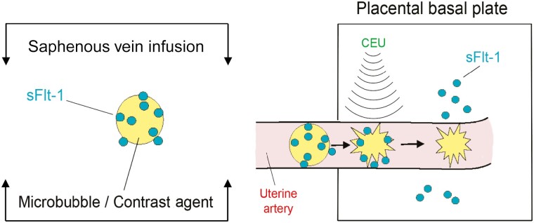
Uterine spiral artery remodeling (SAR) is essential for promoting placental perfusion and fetal development. A defect in SAR results in placental ischemia and increase in placental expression and serum levels of the soluble fms-like tyrosine kinase-1 (sFlt-1) receptor that binds to and suppresses vascular endothelial growth factor (VEGF) bioavailability, thereby leading to maternal vascular dysfunction. We have established a nonhuman primate model of impaired SAR and maternal vascular dysfunction by prematurely elevating estradiol levels in early baboon pregnancy. However, it is unknown whether this primate model of defective SAR involves an increase in placental expression of sFlt-1, which may suppress VEGF bioavailability and thus SAR in the first trimester. Therefore, to establish the role of sFlt-1 in early pregnancy, SAR was quantified in baboons treated on days 25 through 59 of gestation (term = 184 days) with estradiol or with the sFlt-1 gene targeted selectively to the placental basal plate by ultrasound-mediated/microbubble-facilitated gene delivery technology. Placental basal plate sFlt-1 protein expression was 2-fold higher (P < 0.038) and the level of SAR for vessels > 25 µm in diameter was 72% and 63% lower (P < 0.01), respectively, in estradiol-treated and sFlt-1 gene-treated baboons than in untreated animals. In summary, prematurely elevating estradiol levels or sFlt-1 gene delivery increased placental basal plate sFlt-1 protein expression and suppressed SAR in early baboon pregnancy. This study makes the novel discovery that in elevated levels sFlt-1 has a role both in suppressing SAR in early primate pregnancy and maternal vascular endothelial function in late gestation.

Keywords: 1 primate spiral artery remodeling; sFlt.


Figures

Figure 1.
Figure 1.
Vector map of NTC8685-human sFlt-1-i13 IRES-EGFP. CMV, cytomegalovirus; EGFP, enhanced GFP; HTLV-1, human T-cell leukemia virus type 1; IRES (EMCV), internal ribosomal entry site (from the encephalomyocarditis virus); PAS, primosomal assembly site; PAS-BH, primosomal assembly site sequence on the pBR322 H strand; PAS-BL, primosomal assembly site sequence on the pBR322 L strand; pUC, plasmid University of California; SV, simian virus; trpA, tryptophan A; VA RNAI, virus-associated RNA I, a type of noncoding RNA that plays a role in regulating translation.
Figure 2.
Figure 2.
Illustration of UMGD targeted delivery of the sFlt-1 gene to the placental basal plate during early baboon delivery.
Figure 3.
Figure 3.
(A) Grayscale B-mode image of the endometrium (E), placenta (P), and fetus (F) and UMGD contrast-specific images illustrating localization of the sFlt-1 DNA conjugated MBs within the placental basal plate (B) before and (C) after collapse of the MBs/uncoupling of sFlt-1 DNA on day 35 of gestation in a plasmid sFlt-1-treated baboon.
Figure 4.
Figure 4.
Representative photomicrographs of immunocytochemical localization (3,3′-diaminobenzidine brown precipitate) of sFlt-1 protein in the placental floating villi of an sFlt-1 gene-treated baboon (A), proximal anchoring villi (PAV), distal anchoring villi (DAV), and trophoblastic shell (TS) in untreated (B), estradiol-treated (C), and sFlt-1 gene-treated (D) baboons, nonremodeled spiral arteries (SA) in estradiol-treated (E) and sFlt-1 gene-treated (F) baboons, and extravillous trophoblast (EVT) remodeled SA in estradiol-treated (G) and plasmid sFlt-1 gene-treated (H) baboons on day 60 of gestation. Scale bar, 100 µm in all panels.
Figure 5.
Figure 5.
Representative photomicrographs of the localization of red immunofluorescent sFlt-1 signals by proximity ligation assay in the distal anchoring villi (DAV, panels A-C), trophoblastic shell (TS, panels D-F), a nonremodeled spiral artery (G) and extravillous trophoblast remodeled spiral arteries (SA, panels H and I) on day 60 of gestation in untreated (A, D, G), estradiol-treated (B, E, H), and plasmid sFlt-1 gene-treated (C, F, I) baboons. Extravillous trophoblasts are labeled green (cytokeratin). Nuclei are labeled blue. Scale bar, 25 µm for all images.
Figure 6.
Figure 6.
(A) Mean (± SE) sFlt-1 protein levels (signals/nuclear area × 104) as quantified by proximity ligation assay and IP Lab/MetaMorph software in the placental floating villi (FV), proximal anchoring villi (PAV), distal anchoring villi (DAV), and trophoblastic shell (TS) on day 60 of gestation in baboons untreated (n = 3), treated with estradiol on days 25 to 59 (n = 4), or treated with the sFlt-1 gene as targeted by UMGD to the placental basal plate on days 25, 35, 45, and 55 of gestation (n = 5). (B) Mean (± SE) sFlt-1 protein levels collectively in the proximal anchoring villi, distal anchoring villi, and trophoblastic shell in the untreated, estradiol-treated, and sFlt-1 gene-treated baboons in which individual placental basal plate localization is shown in panel A. Values in bars with different lettering are statistically different from one another (2-way ANOVA and Tukey honestly significant difference test) (P < 0.008-< 0.038).
Figure 7.
Figure 7.
(A) Mean (±SE) percentage of SAR (ie, the number of vessels exhibiting extravillous trophoblast invasion divided by total number of vessels counted) classified by vessel diameters of 26 to 50, 51 to 100, and > 100 µm in baboons untreated (n = 9), treated with estradiol daily on days 25 to 59 of gestation (n = 17), or treated with the sFlt-1 gene targeted by UMGD to the placental basal plate on days 25, 35, 45, and 55 (n = 5). (B) Percentage of extravillous trophoblast remodeling of uterine spiral arteries collectively > 25 µm in diameter on day 60 of gestation. Values of bars with different lettering are statistically different (P < 0.05-< 0.01) from one another (individual 1-way ANOVA and Tukey Kramer multiple test). (C) Correlation coefficient SAR and 95% confidence intervals of percent of uterine spiral arteries (collectively < 25 µm in diameter) and sFlt-1 protein levels (collectively in proximal and distal anchoring villi and trophoblastic shell) in untreated, estradiol-treated, and sFlt-1 gene-treated baboons.
Figure 8.
Figure 8.
Role of elevated levels of placental and serum sFlt-1, as induced by prematurely elevating estrogen or sFlt-1 gene delivery in the present study, in suppressing SAR in the first trimester (red lettering) and maternal vascular function in the third trimester (shown previously; 14-21, 34) of primate pregnancy.

References

    1. Hamilton WJ, Boyd JD. Development of the human placenta in the first three months of gestation. J Anat. 1960;94(Pt 3):297-328. - PMC - PubMed
    1. Ramsey EM, Houston ML, Harris JW. Interactions of the trophoblast and maternal tissues in three closely related primate species. Am J Obstet Gynecol. 1976;124(6):647-652. Doi: 10.1016/0002-9378(76)90068-5 - DOI - PubMed
    1. Pijnenborg R, Dixon G, Robertson WB, Brosens I. Trophoblastic invasion of human decidua from 8 to 18 weeks of pregnancy. Placenta. 1980;1(1):3-19. Doi: 10.1016/s0143-4004(80)80012-9 - DOI - PubMed
    1. Enders AC, King BF. Early stages of trophoblastic invasion of the maternal vascular system during implantation in the macaque and baboon. Am J Anat. 1991;192(4):329-346. Doi: 10.1002/aja.1001920403 - DOI - PubMed
    1. Albrecht ED, Pepe GJ. Regulation of uterine spiral artery remodeling: a review. Reprod Sci. 2020;27(10):1932-1942. Doi: 10.1007/s43032-020-00212-8 - DOI - PMC - PubMed

Publication types

Substances