. 2022 Aug 26;226(3):463-473.
doi: 10.1093/infdis/jiac020.

Cytomegalovirus Latent Infection is Associated with an Increased Risk of COVID-19-Related Hospitalization

Collaborators, Affiliations

Cytomegalovirus Latent Infection is Associated with an Increased Risk of COVID-19-Related Hospitalization

Cécile Alanio et al. J Infect Dis. .

Abstract

Some risk factors for severe coronavirus disease 2019 (COVID-19) have been identified, including age, race, and obesity. However, 20%-50% of severe cases occur in the absence of these factors. Cytomegalovirus (CMV) is a herpesvirus that infects about 50% of all individuals worldwide and is among the most significant nongenetic determinants of immune system. We hypothesized that latent CMV infection might influence the severity of COVID-19. Our analyses demonstrate that CMV seropositivity is associated with more than twice the risk of hospitalization due to severe acute respiratory syndrome coronavirus 2 (SARS-CoV-2) infection. Immune profiling of blood and CMV DNA quantitative polymerase chain reaction in a subset of patients for whom respiratory tract samples were available revealed altered T-cell activation profiles in absence of extensive CMV replication in the upper respiratory tract. These data suggest a potential role for CMV-driven immune perturbations in affecting the outcome of SARS-CoV-2 infection and may have implications for the discrepancies in COVID-19 severity between different human populations.

Keywords: CMV; COVID-19; EMRA T cells; immune profiling; risk factor.


Figures

Figure 1.
Figure 1.
Cytomegalovirus (CMV) seropositivity is associated with an increased risk of testing positive for severe acute respiratory syndrome coronavirus 2 (SARS-CoV-2). A, Mosaic plot showing the numbers and proportions of individuals with SARS-CoV-2-positive or SARS-CoV-2–negative reverse-transcription polymerase chain reaction results as a function of CMV serostatus. B, Odds ratios and 95% confidence intervals for testing positive for SARS-CoV-2 (sample size, n = 984); red indicates a P value <.05, as indicated in Table 1.
Figure 2.
Figure 2.
Cytomegalovirus (CMV) seropositivity is associated with an increased risk of hospitalization related to coronavirus disease 2019 (COVID-19). A, Frequency of CMV seropositivity or seronegativity among individuals testing positive for COVID-19, including those not hospitalized (group [Gp] 1; n = 167), those hospitalized but not in the intensive care unit (ICU) (Gp 2; n = 49), and those hospitalized in the ICU (Gp 3; n = 30). P value is based on binomial logistic regression including patient age, sex, and self-reported race. B, Mosaic plot showing the numbers and proportions of hospitalizations due to COVID-19 as a function of CMV serostatus (sample size, n = 246). C, Odds ratios and 95% confidence intervals for hospitalization related to COVID-19 (sample size, n = 246); red indicates a P value <.05, as indicated in Table 2. D, Mean CMV immunoglobulin (Ig) G levels (in standard units [SU]) and standard deviations (SDs) as a function of COVID-19 severity in CMV-seropositive individuals (sample size, n = 246).
Figure 3.
Figure 3.
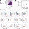
Cytomegalovirus (CMV) reactivation and effector memory reexpressing CD45RA (EMRA) T-cell activation in patients with coronavirus disease 2019 (COVID-19). A, Frequency of CMV seropositivity and seronegativity among patients recovered from mild COVID-10 (n = 27), healthy controls (n = 24), patients with COVID-19 (n = 127), and patients with non–COVID-19 acute respiratory distress syndrome (ARDS) (n = 44). Of note, these models were adjusted by age and sex but not by self-reported race, as this information was not available for healthy donors. B, CMV DNA quantitative polymerase chain reaction (qPCR) as a function of CMV serostatus, in respiratory tract samples from 40 patients with COVID-19. C, Longitudinal assessment of proportions of EMRA CD8+ T cells (red squares) using CD45RA/CD27, as well as Ki67+ and CD38+HLA-DR+ EMRA CD8+ T cells in 2 patients with CMV reactivation in the upper respiratory tract. The CMV DNA qPCR status at the time of sampling is indicated. Abbreviation: FSC-W, forward scatter width; Pt, patient.
Figure 4.
Figure 4.
Cytomegalovirus (CMV) seropositivity in patients with coronavirus disease 2019 (COVID-19) is associated with effector memory reexpressing CD45RA (EMRA) T-cell activation. A, Quantification of the effect of CMV seropositivity on the abundance of circulating immune cells obtained from donors with COVID-19 (n = 127), estimated in a linear mixed model with a log-transformed immunophenotype as the response, controlled for age, sex, and self-reported race, then transformed to the original scale (with 99% confidence intervals [CIs] adjusted for false coverage). Abbreviations: cTfh, conventional T follicular helper cell; EM1, effector memory 1; PD-1, programmed cell death 1 protein. B, Unmodified Uniform Manifold Approximation and Projection (UMAP) projection of patients recovered from COVID-19 (n = 27), healthy controls (n = 24), and patients with COVID-19 (n = 127), as described elsewhere [15], using single-positive populations of all immune cell subsets. Individuals are color coded by their CMV serostatus; black square indicates cluster of CMV-seropositive patients (n = 20) further examined in D. Abbreviation: NA, non available. C, UMAP model illustrated as described elsewhere [15], with feature-weighted density contours overlaid on patients with COVID-19 (n = 127). The illustrated gradient vector was computed according to the direction of maximum Pearson correlation for the flow cytometric feature of interest (here, EMRA CD8+ T cells) and weighted in length according to the strength of the resulting Pearson coefficient. Red lines indicate high EMRA; blue lines, low EMRA T-cell proportions. The intensity of the dots represents the z score, and the dotted line is drawn perpendicular to the gradient vector. Triangles represent deaths within 90 days of admission, as reported elsewhere [15]. D, Immunophenotypes (n = 279) significantly associated with the cluster of CMV-seropositive patients (sample size, n = 20) displayed in B, as estimated in a linear mixed model with a log-transformed immunophenotype as the response, controlled for age, sex, and self-reported race and then transformed to the original scale (with 99% CIs adjusted for false coverage).
Figure 5.
Figure 5.
A portion of effector memory reexpressing CD45RA (EMRA) CD8+ T cells activated during coronavirus disease 2019 (COVID-19) are cytomegalovirus (CMV) specific. A, Identification of interferon (IFN) γ+41BB+CD8+ T cells within CD45RA+CD27+CCR7+ (naive), CD45RACD27+CCR7+ (central memory), CD45RACD27+CCR7 (effector memory 1), CD45RACD27CCR7 (effector memory 2), CD45RACD27CCR7+ (effector memory 3), CD45RA+CD27 (EMRA) [15] in an unstimulated (top row) and a CMV peptide–stimulated (bottom row) donor. B, Overlay of IFN-γ+41BB+CD8+ T cells on a CD45RA/CD27 scatterplot from the donor as in A. C, Summary of frequencies of IFN-γ+41BB+CD8+ T cells within T-cell subsets as in A, in 4 unstimulated (blue dots) and CMV-peptide stimulated (red dots) donors.

References

    1. World Health Organization. Coronavirus disease (COVID-19) weekly epidemiological update and weekly operational update. 2021:1–25. Available at: https://www.who.int/emergencies/diseases/novel-coronavirus-2019/situatio...
    1. Huang C, Wang Y, X Li, et al. . Clinical features of patients infected with 2019 novel coronavirus in Wuhan, China. Lancet 2020; 395:497–506. - PMC - PubMed
    1. Grasselli G, Tonetti T, Filippini C, Slutsky AS, Pesenti A, Ranieri VM.. Pathophysiology of COVID-19-associated acute respiratory distress syndrome. Lancet Respir Med 2021; 9:e5–6. - PMC - PubMed
    1. Zhou F, T Y, R D, et al. . Clinical course and risk factors for mortality of adult inpatients with COVID-19 in Wuhan, China: a retrospective cohort study. Lancet 2020; 395:1054–62. - PMC - PubMed
    1. Wu JT, Leung K, Bushman M, et al. . Estimating clinical severity of COVID-19 from the transmission dynamics in Wuhan, China. Nat Med 2020; 1:13. - PMC - PubMed

Publication types