Review
. 2022 Apr 26:40:615-649.
doi: 10.1146/annurev-immunol-101220-014952. Epub 2022 Feb 8.

Distinct Cellular Tropism and Immune Responses to Alphavirus Infection

Affiliations
Review

Distinct Cellular Tropism and Immune Responses to Alphavirus Infection

Natasha M Kafai et al. Annu Rev Immunol. .

Abstract

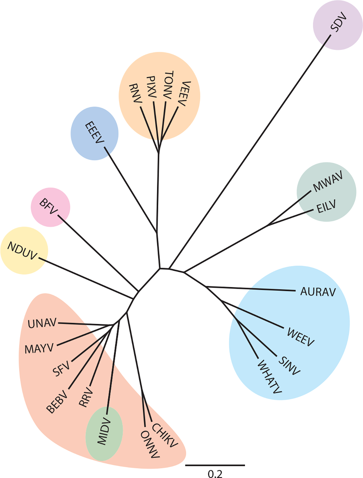
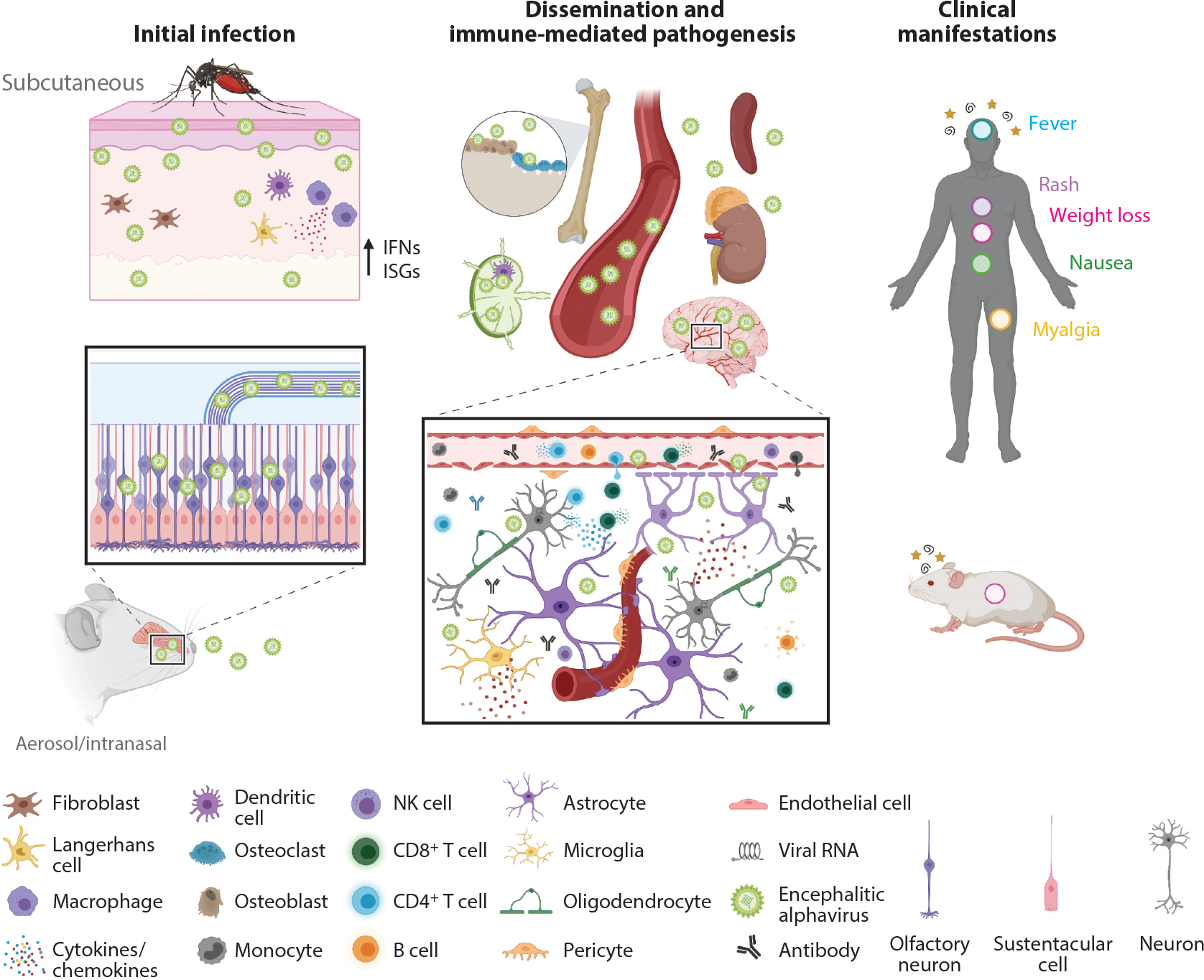
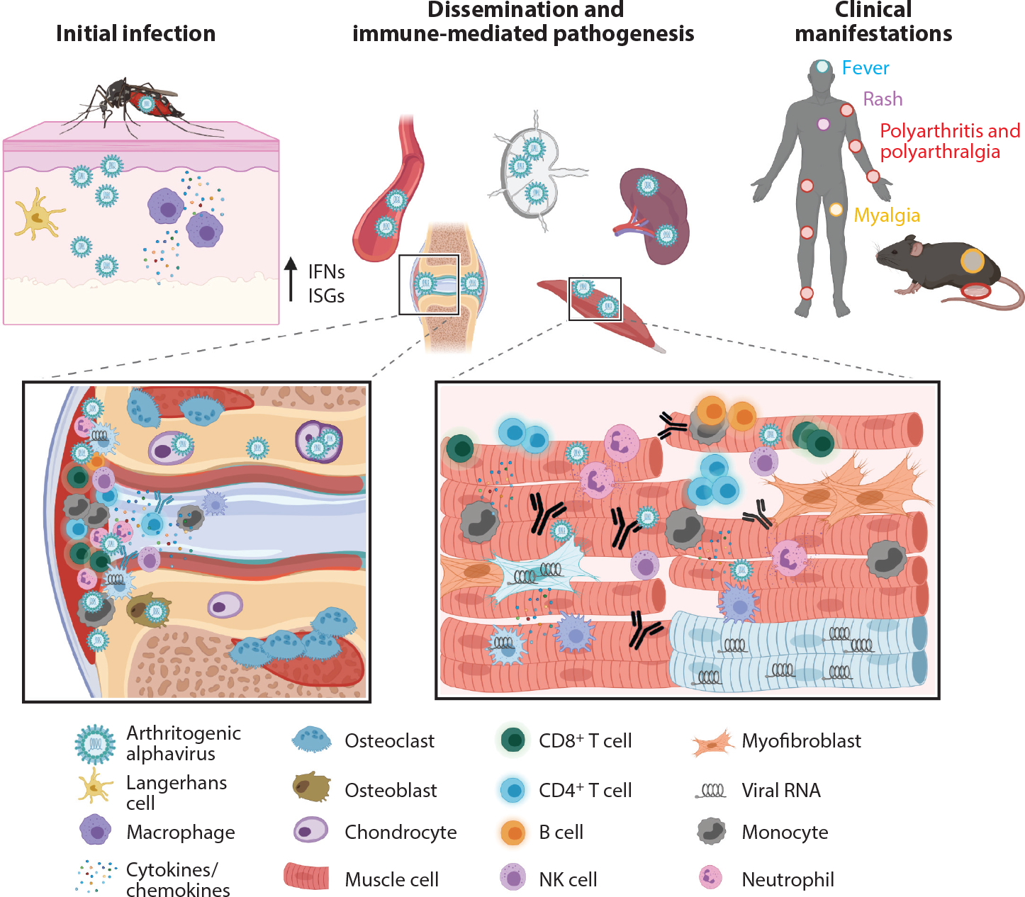
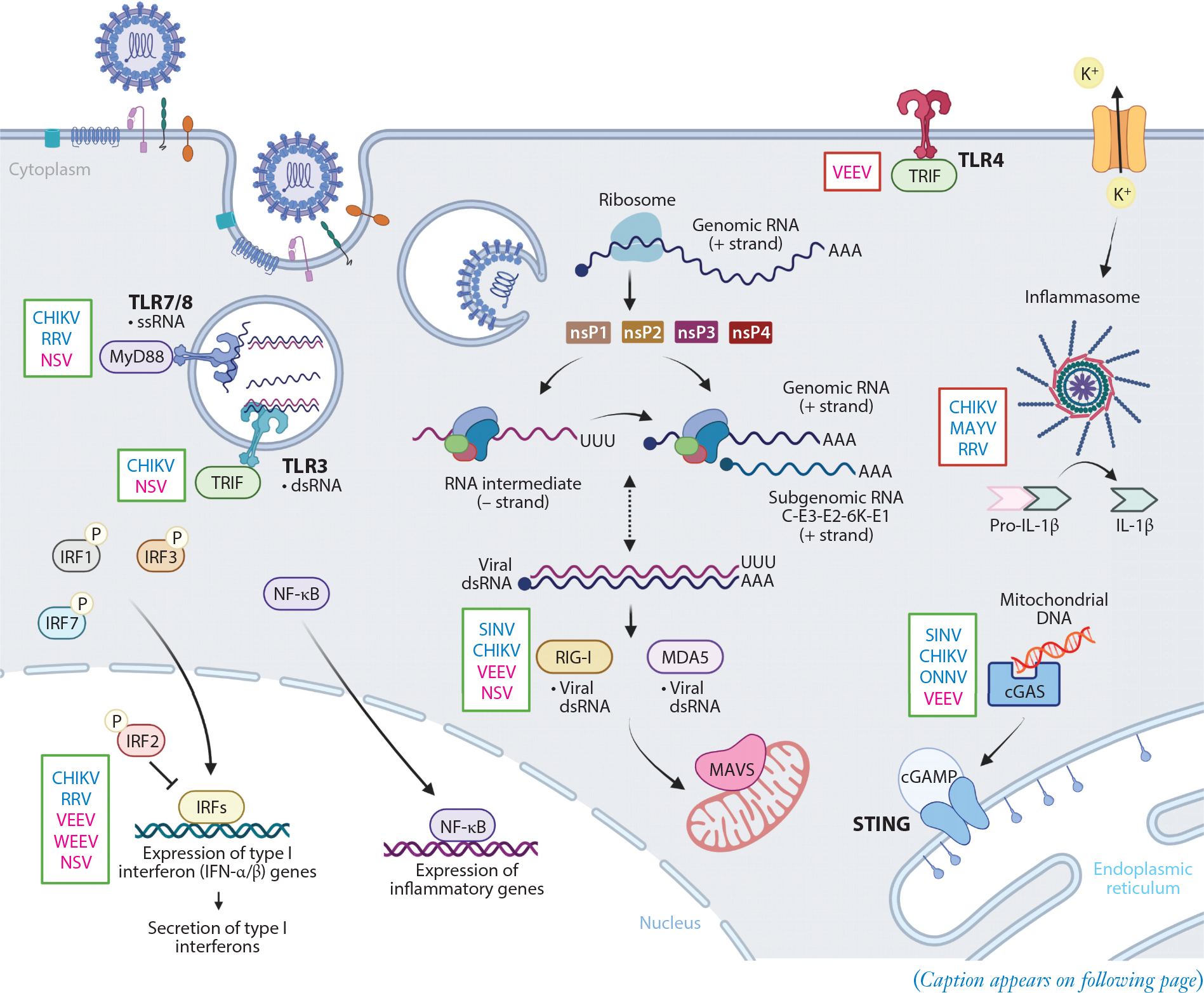
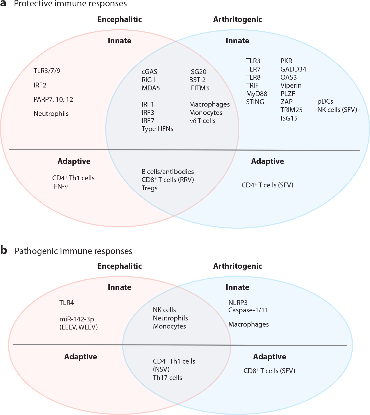
Alphaviruses are emerging and reemerging viruses that cause disease syndromes ranging from incapacitating arthritis to potentially fatal encephalitis. While infection by arthritogenic and encephalitic alphaviruses results in distinct clinical manifestations, both virus groups induce robust innate and adaptive immune responses. However, differences in cellular tropism, type I interferon induction, immune cell recruitment, and B and T cell responses result in differential disease progression and outcome. In this review, we discuss aspects of immune responses that contribute to protective or pathogenic outcomes after alphavirus infection.

Keywords: alphavirus; arbovirus; immunity; mouse models; pathogenesis; tropism.


Figures

Figure 1
Figure 1
Genetic relationships of alphaviruses. Aquatic alphavirus (purple): sleeping disease virus (SDV) (accession number Q8QL52.1). Insect-only alphaviruses (teal): Mwinilunga virus (MWAV) (BBC45635) and Eilat virus (EILV) (QBG67157). Western equine encephalitis virus complex (cyan): Aura virus (AURAV) (Q86925), western equine encephalitis virus (WEEV) (QEX51909.1), Sindbis virus (SINV) (AAM10630.1), and Whataroa virus (WHATV) (AAO33329). Semliki Forest virus complex (red): chikungunya virus (CHIKV) (ABO38821), o’nyong-nyong virus (ONNV) (AAC97205), Ross River virus (RRV) (AYI50356.1), bebaru virus (BEBV) (AEJ36225), Semliki Forest virus (SFV) (QQZ00842), Mayaro virus (MAYV) (QDL88200.1), and Una virus (UNAV) (YP_009665989). Middelburg virus complex (green): Middelburg virus (MIDV) (AAO33343). Ndumu virus complex (yellow): Ndumu virus (NDUV) (AAO33345). Barmah Forest virus complex (pink): Barmah Forest virus (BFV) (AAO33347). Eastern equine encephalitis virus complex (dark blue): eastern equine encephalitis virus (EEEV) (AMT80322.1). Venezuelan equine encephalitis virus complex (orange): Rio Negro virus (RNV) (YP_009507803), Pixuna virus (PIXV) (YP_009507801), Tonate virus (TONV) (AAD14557), and Venezuelan equine encephalitis virus (VEEV) (AAC19322.1). The C-E3-E2–6K-E1 amino acid sequences were aligned using Geneious Aligner, and a phylogenic tree was built using Geneious Tree Builder.
Figure 2
Figure 2
Infection and pathogenesis of encephalitic alphaviruses. Inoculation by encephalitic alphaviruses can occur subcutaneously through the bite of a mosquito or through an intranasal or aerosolized exposure. In the skin, some encephalitic alphaviruses (e.g., VEEV) will replicate locally in dendritic cells and macrophages, prompting release of type I interferons and expression of ISGs. Virus then travels to the draining lymph node or through the bloodstream to disseminate to peripheral tissues. Other encephalitic alphaviruses, like EEEV, replicate in fibroblasts and osteoblasts but not in lymphoid tissues and do not induce a measurable peripheral inflammatory response. Virus spreads rapidly to the CNS via several routes, including retrograde transport in neurons of the olfactory bulb and trigeminal nerves, hematogenous routes, and across the BBB. Release of proinflammatory cytokines and chemokines alters and compromises the BBB, contributing to further CNS infection. Immune cells are recruited into the brain parenchyma, and resident glia become activated, both of which are essential for control of infection. Nonetheless, activated immune cells can also damage neurons and cause demyelination, which drives the neurological syndromes observed in humans (stars and spirals). Depending on the virus strain, neurons and glia can survive infection and harbor viral RNA after acute infection. Other signs and symptoms of systemic disease include fever (blue circle), rash (purple circle), weight loss (pink circle), nausea (green circle), and myalgia (yellow circle). Abbreviations: BBB, blood-brain barrier; CNS, central nervous system; EEEV, eastern equine encephalitis virus; IFN, interferon; ISG, interferon-stimulated gene; NK, natural killer; VEEV, Venezuelan equine encephalitis virus. Figure adapted from images created with BioRender.com.
Figure 3
Figure 3
Infection and pathogenesis of arthritogenic alphaviruses. Following a bite from an infected mosquito, arthritogenic alphaviruses replicate locally in fibroblasts, keratinocytes, epithelial and endothelial cells, and/or macrophages. Cellular infection results in induction of type I interferons and expression of ISGs. Virus travels to the lymph node or through the bloodstream to disseminate to peripheral organs, where it replicates to high titers in the joint-associated and musculoskeletal tissues. Infection of osteoblasts promotes osteoclastogenesis and bone erosion. Proinflammatory cytokines and chemokines are released and recruit cells that are essential to control infection but also mediate damage including synovitis, bone reabsorption, and muscle fiber destruction. Macrophages, myofibroblasts, and muscle cells can survive infection and harbor viral RNA many weeks after acute infection. These processes drive the clinical syndromes observed in humans, including fever (blue circle), rash (purple circle), polyarthritis and polyarthralgia (red circles), and myalgia (yellow circle). Abbreviations: IFN, interferon; ISG, interferon-stimulated gene; NK, natural killer. Created with BioRender.com.
Figure 4
Figure 4
Overview of innate immune signaling pathways impacting alphavirus infection. Alphavirus entry is mediated by attachment factors and receptors at the host cell surface. Once internalized in an endosome, alphaviruses undergo fusion of the viral envelope with the endosomal membrane to release nucleocapsid and positive-strand viral genomic RNA into the cytoplasm. This RNA is translated to generate nonstructural proteins (nsP1–4) that form an initial viral replication complex to synthesize a negative-strand RNA intermediate. The viral replication complex will then switch to generate positive-strand genomic RNA and (positive-strand) subgenomic RNA from these negative-strand RNA intermediates. Subgenomic RNA encodes the structural polyproteins (C-E3-E2–6K-E1), and these are translated and processed in the endoplasmic reticulum before delivery to the cell surface to encapsulate genomic RNA during virion budding. PRRs at the cell surface (TLR4) and within the endosome (TLR3, TLR7, and TLR8) will recognize PAMPs and signal through distinct adaptor proteins (e.g., MyD88 and TRIF). TLR activation, along with secondary signaling through potassium efflux, can activate the inflammasome. Transcription of viral RNA in the cytoplasm leads to formation of dsRNA, which can activate cytoplasmic PRRs, RIG-I and MDA5, to interact with MAVS on the mitochondrial membrane. cGAS can recognize endogenous or foreign DNA released during infection, leading to activation of STING. These recognition events initiate translocation of IRFs and NF-κB into the nucleus and regulation of type I interferons and proinflammatory gene expression. Innate immune pathways that confer presumed protective (green box) or pathological (red box) responses to arthritogenic (blue font) or encephalitic (pink font) alphavirus infection are indicated to the left of each represented signaling molecule. Abbreviations: cGAMP, cyclic GMP-AMP; cGAS, cGAMP synthase; CHIKV, chikungunya virus; dsRNA, double-stranded RNA; IRF, interferon regulatory factor; MAVS, mitochondrial antiviral-signaling protein; MAYV, Mayaro virus; NF-κB, nuclear factor kappa B; NSV, neuroadapted SINV; ONNV, o’nyong-nyong virus; PAMP, pathogen-associated molecular pattern; PRR, pattern recognition receptor; RRV, Ross River virus; SINV, Sindbis virus; ssRNA, single-stranded RNA; STING, stimulator of interferon genes; TLR, Toll-like receptor, VEEV, Venezuelan equine encephalitis virus; WEEV, western equine encephalitis virus. Adapted from images created with BioRender.com.
Figure 5
Figure 5
Protective and pathogenic components of the immune response to alphavirus infections. Pattern recognition receptors, type I interferons, interferon-stimulated genes, and immune cells involved in the innate and adaptive immune responses to alphaviruses that are encephalitic (pink ovals), arthritogenic (light blue ovals), or both encephalitic and arthritogenic (mauve overlap) are categorized by their known or presumed (a) protective and/or (b) pathogenic contributions. Specific viruses are indicated when the type of immune response differs compared to other viruses in the respective group. Abbreviations: BST-2, bone marrow stromal cell antigen 2; cGAS, cyclic GMP-AMP synthase; EEEV, eastern equine encephalitis virus; GADD34, growth arrest and DNA damage–inducible protein 34; IFITM3, interferon-induced transmembrane 3; IRF1, interferon regulatory factor 1; ISG20, interferon-stimulated gene 20; NK, natural killer; NSV, neuroadapted Sindbis virus; OAS3, 2′,5′-oligoadenylate synthetase 3; PARP7, poly(ADP-ribose) polymerase 7; pDC, plasmacytoid dendritic cell; PKR, protein kinase R; PLZF, promyelocytic leukemia zinc finger; RRV, Ross River virus; SFV, Semliki Forest virus; STING, stimulator of interferon genes; Th1, T helper type 1; TLR3, Toll-like receptor 3; Treg, regulatory T cell; TRIM25, tripartite motif-containing 25; WEEV, western equine encephalitis virus; ZAP, zinc finger antiviral protein.

Similar articles

Cited by

References

    1. Weaver SC, Barrett AD. 2004. Transmission cycles, host range, evolution and emergence of arboviral disease. Nat. Rev. Microbiol. 2:789–801 - PMC - PubMed
    1. Leung JY, Ng MM, Chu JJ. 2011. Replication of alphaviruses: a review on the entry process of alphaviruses into cells. Adv. Virol. 2011:249640. - PMC - PubMed
    1. Li L, Jose J, Xiang Y, Kuhn RJ, Rossmann MG. 2010. Structural changes of envelope proteins during alphavirus fusion. Nature 468:705–8 - PMC - PubMed
    1. Weaver SC, Winegar R, Manger ID, Forrester NL. 2012. Alphaviruses: population genetics and determinants of emergence. Antivir. Res. 94:242–57 - PMC - PubMed
    1. Levi LI, Vignuzzi M. 2019. Arthritogenic alphaviruses: a worldwide emerging threat? Microorganisms 7:133. - PMC - PubMed

Publication types

Substances

LinkOut - more resources