. 2022 Feb 8;13(1):66.
doi: 10.1186/s13287-022-02717-2.

Synergistic regenerative therapy of thin endometrium by human placenta-derived mesenchymal stem cells encapsulated within hyaluronic acid hydrogels

Affiliations

Synergistic regenerative therapy of thin endometrium by human placenta-derived mesenchymal stem cells encapsulated within hyaluronic acid hydrogels

Yifeng Lin et al. Stem Cell Res Ther. .

Abstract

Background: Thin endometrium is a primary cause of defective endometrial receptivity, resulting in infertility or recurrent miscarriage. Much effort has been devoted toward regenerating thin endometrium by stem cell-based therapies. The human placenta-derived mesenchymal stem cells (HP-MSCs) are emerging alternative sources of MSCs with various advantages. To maximize their retention inside the uterus, we loaded HP-MSCs with cross-linked hyaluronic acid hydrogel (HA hydrogel) to investigate their therapeutic efficacy and possible underlying mechanisms.

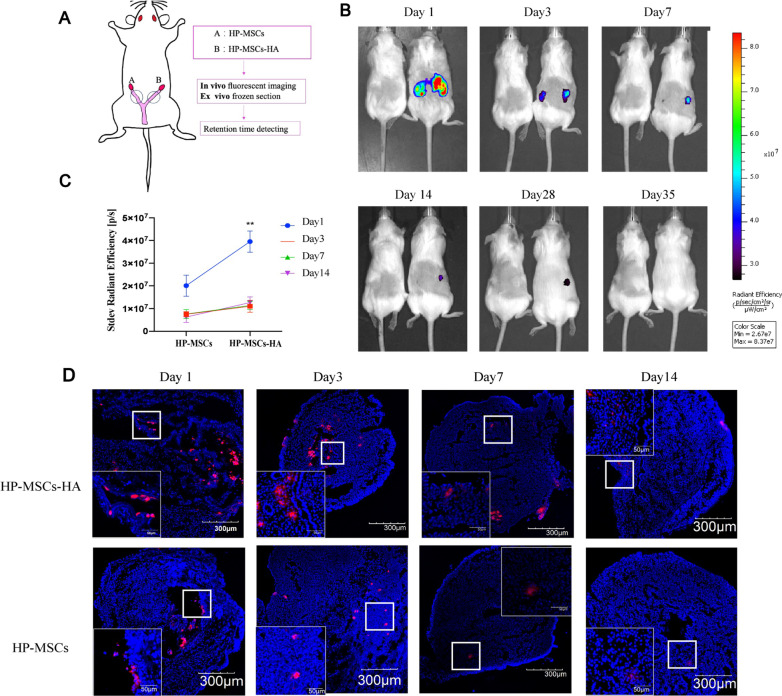
Methods: Ethanol was injected into the mice uterus to establish the endometrium-injured model. The retention time of HP-MSCs and HA hydrogel was detected by in vivo imaging, while the distribution of HP-MSCs was detected by immunofluorescence staining. Functional restoration of the uterus was assessed by testing embryo implantation rates. The endometrial morphological alteration was observed by H&E staining, Masson staining, and immunohistochemistry. In vitro studies were further conducted using EdU, transwell, tube formation, and western blot assays.

Results: Instilled HP-MSCs with HA hydrogel (HP-MSCs-HA) exhibited a prolonged retention time in mouse uteri than normal HP-MSCs. In vivo studies showed that the HP-MSCs-HA could significantly increase the gland number and endometrial thickness (P < 0.001, P < 0.05), decrease fibrous area (P < 0.0001), and promote the proliferation and angiogenesis of endometrial cells (as indicated by Ki67 and VEGF, P < 0.05, P < 0.05, respectively) in mice injured endometrium. HP-MSCs-HA could also significantly improve the embryo implantation rate (P < 0.01) compared with the ethanol group. Further mechanistic study showed the paracrine effects of HP-MSCs. They could not only promote the proliferation and migration of human endometrial stromal cells via the JNK/Erk1/2-Stat3-VEGF pathway but also facilitate the proliferation of glandular cells via Jak2-Stat5 and c-Fos-VEGF pathway. In turn, the increased VEGF in the endometrium promoted the angiogenesis of endothelial cells.

Conclusion: Our study suggested the potential therapeutic effects and the underlying mechanisms of HP-MSCs-HA on treating thin endometrium. HA hydrogel could be a preferable delivery method for HP-MSCs, and the strategy represents a promising therapeutic approach against endometrial injury in clinical settings.

Keywords: Endometrial repair; Human placenta-derived mesenchymal stem cells; Hyaluronic acid hydrogels; Regeneration mechanisms; Thin endometrium.


Conflict of interest statement

The authors declare that they have no competing interests.

Figures

Fig. 1
Fig. 1
Fabrication and characterization of HA hydrogel. A The synthesis routine of GM-HA. B The 1H-NMR spectrum of GM-HA in D2O. C Schematic presentation of preparation procedures of HA hydrogel and HP-MSCs-HA as well as the instillation of HP-MSCs-HA into a mouse model. D The FT-IR spectra of GM-HA and HA hydrogel. E The storage modulus G’ and loss modulus G’’ of HA hydrogel versus oscillation frequency from 0.01 to 100 rad/s with a fixed stain of 0.2% at 37 °C. F SEM morphology of freeze-dried HA. G SEM morphology of HA hydrogel. The scale bars in (F) and (G) were 100 μm
Fig. 2
Fig. 2
The retention time and distribution of HP-MSCs in the endometrium. A Schematic presentation of intrauterine instillation and its corresponding detection methods. Equal cell quantities of HP-MSCs and HP-MSCs-HA labelled by CM-DiD or CM-DiR were implanted to the certain uterus (2 × 105 per/uterus). B The mouse on the left was the untreated control group to eliminate the interference of autofluorescence, while the mouse on the right was the treatment group. The retention time of HP-MSCs was observed by an IVIS Spectrum Imaging System at the different time points (1, 3, 7, 14, 28, and 35 days) after transplantation. C The total radiant efficiency of the treated uterus was measured and calculated. Data were presented as mean ± SEM (n = 3). ** indicates P < 0.01. D Ex vivo frozen section of the uterus showed the retention of HP-MSCs, which were labelled with CM-DiD (red). The nuclei of cells were labelled with DAPI (blue)
Fig. 3
Fig. 3
The construction of endometrium-injured mouse model and the evaluation of embryo implantation after therapy. A Schematic diagram of mice grouping and their detection methods. B, C Evaluate the endometrial receptivity of the five mouse groups with different treatments by the number of implanted embryos. * indicates P < 0.05, ** indicates P < 0.01, **** indicates P < 0.0001, n = 8
Fig. 4
Fig. 4
The histological analysis of endometrium for the five mouse groups after different treatments. A H&E staining results of the five groups for evaluating the endometrial thickness and number of glands. B Masson staining results of the five groups for evaluating the fibrosis status of the endometrium. C Immunohistochemical Ki67 expression of the five groups for evaluating the proliferation of endometrial cells. D Immunohistochemical VEGF expression of the five groups for evaluating the angiogenesis of endometrium. E Average endometrial thickness and statistical analysis (± SEM) of the five groups. * indicates P < 0.05, ** indicates P < 0.01, *** indicates P < 0.001, n = 6. F Average gland number and statistical analysis (± SEM) of the five groups. * indicates P < 0.05, n = 6. G Average fibrosis area and statistical analysis (± SEM) of the five groups. The ratio of the fibrotic area = endometrial fibrotic area/endometrial area. * indicates P < 0.05, ** indicates P < 0.01, *** indicates P < 0.001, **** indicates P < 0.0001, n = 6. H Statistic analysis of IRS of Ki67 in the endometrium of the five groups. * indicates P < 0.05, ** indicates P < 0.01, n = 6. I Statistic analysis of IRS of VEGF in the endometrium of the five groups. * indicates P < 0.05, ** indicates P < 0.01, n = 6
Fig. 5
Fig. 5
Effects of HP-MSCs on the proliferation of human endometrial stromal cells. EdU assays were conducted at 24 h, 48 h, and 72 h, respectively, after co-culturing of human stromal cells with (Ctr) and with HP-MSCs, respectively. AC Representative confocal images of human stromal cells stained with EdU (red) and DAPI (blue) at 24 h, 48 h, and 72 h, respectively. EdU represents the positive proliferation cells, while DAPI represents the cell nucleus. D Schematic diagram of co-culture, HP-MSCs were inoculated in the upper insert transwell (0.4um), while stromal cells were in the lower hole plate. EG EdU/DAPI represented the ratio of proliferating cells to total cells, and the data were shown as mean ± SEM. * indicates P < 0.05, ** indicates P < 0.01, *** indicates P < 0.001
Fig. 6
Fig. 6
Effect of HP-MSCs on the migration of human endometrial stromal cells. A Schematic of co-culturing the stromal cells with HP-MSCs. Stromal cells were inoculated in the upper insert transwell (8 um), while HP-MSCs were in the lower hole plate. B Transwell migration assay was conducted at 24 h, 48 h, and 72 h, respectively, after culturing without and with HP-MSCs, respectively. The migrated cells were stained with purple. C The average number of migrated stromal cells after culturing without or with HP-MSCs for 24 h, 48 h, and 72 h, respectively (± SEM). * indicates P < 0.05, ** indicates P < 0.01, *** indicates P < 0.001. D p-JNK, p-Stat3, p-Erk1/2, VEGF, and corresponding total protein western blot analysis at 24 h, 48 h, and 72 h, respectively, after culturing without and with HP-MSCs, respectively
Fig. 7
Fig. 7
Effect of HP-MSCs on the proliferation of human endometrial glandular cells. AC EdU assays were conducted after co-culturing the glandular cells without and with HP-MSCs for 24 h, 48 h, 72 h, respectively. Representative confocal images of the glandular cells stained with EdU (red) and DAPI (blue). D Schematic of co-culturing the glandular cells with HP-MSCs. HP-MSCs were inoculated in the upper insert transwell (0.4um), while the glandular cells were in the bottom plate. EG Average EdU/DAPI ratio (± SEM). * indicates P < 0.05. H p-Jak2, p-Stat5, p–c-Fos, p–c-Jun, VEGF, and corresponding total protein western blot analysis at 24 h, 48 h, and 72 h, after culturing without and with HP-MSCs, respectively
Fig. 8
Fig. 8
HUVECs angiogenesis assay. A Schematic of co-culturing the HP-MSCs with HUVECs. After 24 h co-culturing with HP-MSCs, HUVECs were seeded in Matrigel-coated u-slides to conduct tube formation assay. B Representative tube formation images of Ctr and HP-MSCs group. C, D Quantitative assay of tube formation assay and data were expressed as mean ± SEM. * indicates P < 0.05

References

    1. Ombelet W, Cooke I, Dyer S, Serour G, Devroey P. Infertility and the provision of infertility medical services in developing countries. Hum Reprod Update. 2008;14(6):605–621. - PMC - PubMed
    1. Carson S, Kallen A. Diagnosis and Management of Infertility: a Review. JAMA. 2021;326(1):65–76. - PMC - PubMed
    1. Lédée-Bataille N, Laprée-Delage G, Taupin JL, Dubanchet S, Frydman R, Chaouat G. Concentration of leukaemia inhibitory factor (LIF) in uterine flushing fluid is highly predictive of embryo implantation. Hum Reprod. 2002;17(1):213–218. - PubMed
    1. Deans R, Abbott J. Review of intrauterine adhesions. J Minim Invasive Gynecol. 2010;17(5):555–569. - PubMed
    1. Cenksoy P, Ficicioglu C, Yıldırım G, Yesiladali M. Hysteroscopic findings in women with recurrent IVF failures and the effect of correction of hysteroscopic findings on subsequent pregnancy rates. Arch Gynecol Obstet. 2013;287(2):357–360. - PubMed

Publication types