. 2022 Apr 1;82(7):1267-1282.
doi: 10.1158/0008-5472.CAN-21-0914.

Lactate Rewires Lipid Metabolism and Sustains a Metabolic-Epigenetic Axis in Prostate Cancer

Affiliations

Lactate Rewires Lipid Metabolism and Sustains a Metabolic-Epigenetic Axis in Prostate Cancer

Luigi Ippolito et al. Cancer Res. .

Abstract

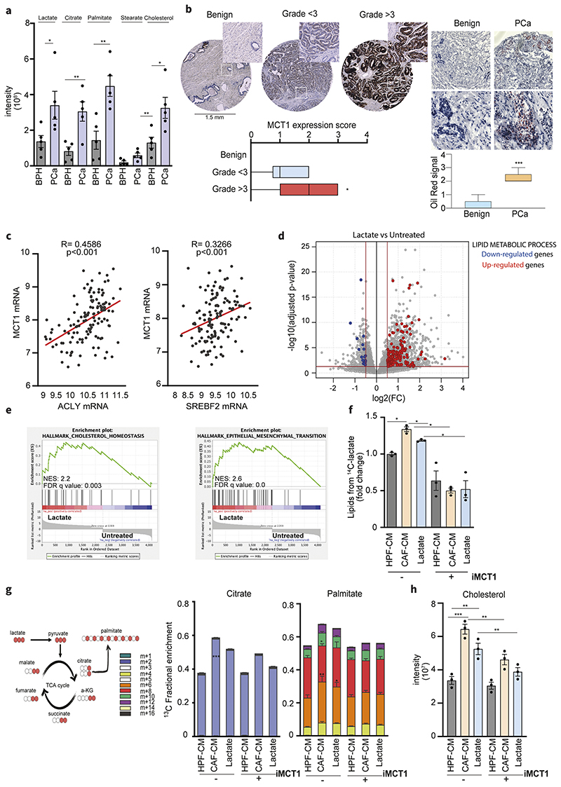
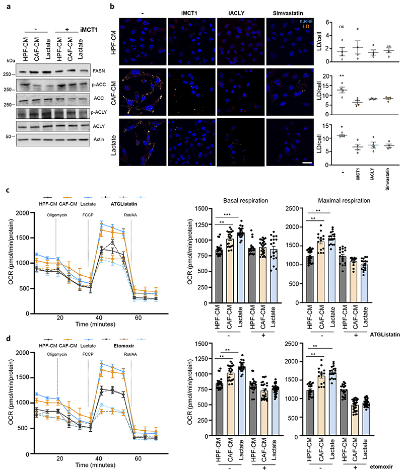
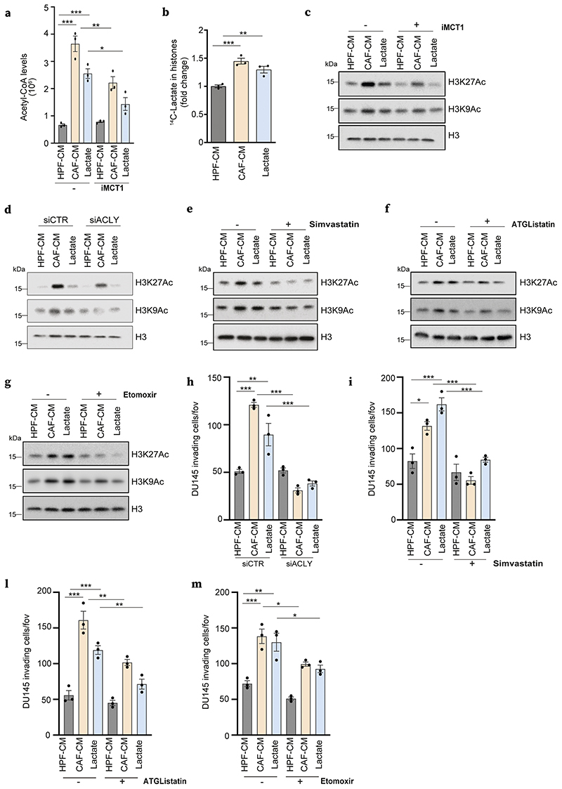
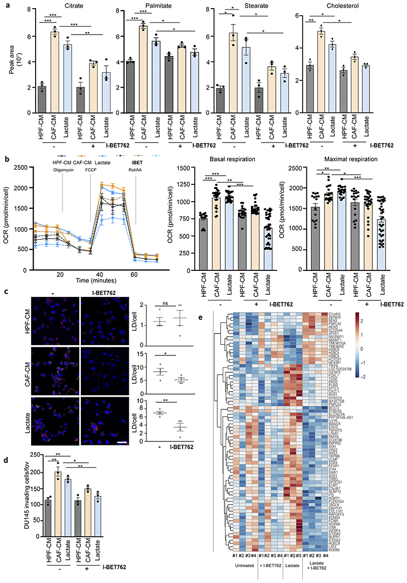
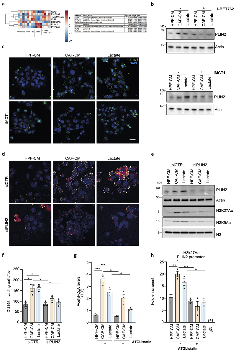
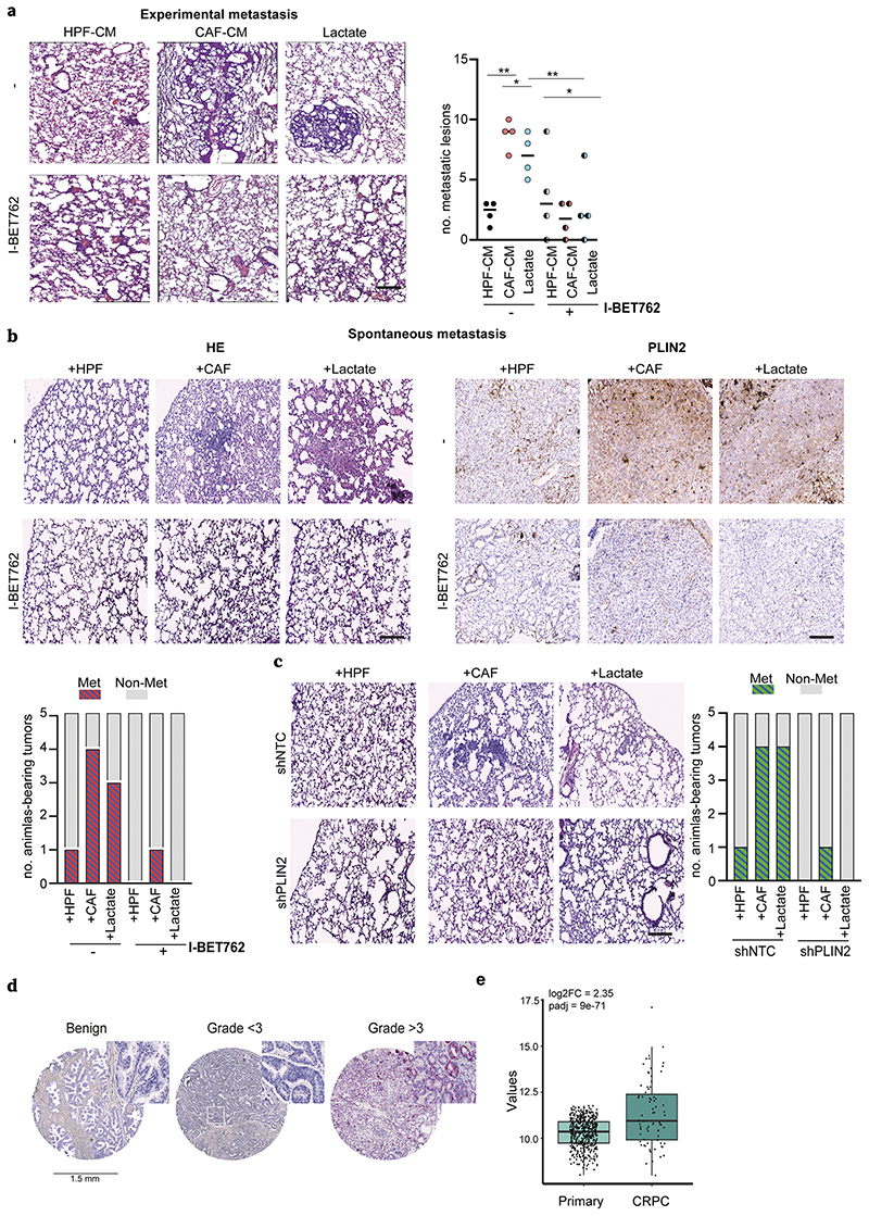
Lactate is an abundant oncometabolite in the tumor environment. In prostate cancer, cancer-associated fibroblasts (CAF) are major contributors of secreted lactate, which can be taken up by cancer cells to sustain mitochondrial metabolism. However, how lactate impacts transcriptional regulation in tumors has yet to be fully elucidated. Here, we describe a mechanism by which CAF-secreted lactate is able to increase the expression of genes involved in lipid metabolism in prostate cancer cells. This regulation enhanced intracellular lipid accumulation in lipid droplets (LD) and provided acetyl moieties for histone acetylation, establishing a regulatory loop between metabolites and epigenetic modification. Inhibition of this loop by targeting the bromodomain and extraterminal protein family of histone acetylation readers suppressed the expression of perilipin 2 (PLIN2), a crucial component of LDs, disrupting lactate-dependent lipid metabolic rewiring. Inhibition of this CAF-induced metabolic-epigenetic regulatory loop in vivo reduced growth and metastasis of prostate cancer cells, demonstrating its translational relevance as a therapeutic target in prostate cancer. Clinically, PLIN2 expression was elevated in tumors with a higher Gleason grade and in castration-resistant prostate cancer compared with primary prostate cancer. Overall, these findings show that lactate has both a metabolic and an epigenetic role in promoting prostate cancer progression.

Significance: This work shows that stromal-derived lactate induces accumulation of lipid droplets, stimulates epigenetic rewiring, and fosters metastatic potential in prostate cancer.


Conflict of interest statement

Conflict of interest: The authors declare no potential conflicts of interest.

Figures

Figure 1
Figure 1. Lactate reprograms lipid metabolism in PCa
a) Levels of lactate, citrate, palmitate, stearate and cholesterol were measured in prostate carcinomas (PCa) and benign hyperplasia (BPH) (n=5). Data represent means ± SEMs. Student’s t test; *p < 0.05; **p < 0.01; ***p < 0.001 b) MCT1 expression in representative tissue cores from TMA (n=63, magnification 20X) including benign and tumor tissue samples with low (< 3) and high Gleason grade (≥3). Box-and-whiskers plots of MCT1 histoscores in tissue samples compared by Gleason grade. On the right, Oil Red staining and quantification in representative PCa and benign tissues (n=5, scale bar: 50μm, magnification 40X). c) Correlation analysis between ACLY/SREBF2 and MCT1 expression in primary tumor specimens of Taylor dataset. Each dot corresponds to an individual specimen. Pearson correlation r. d) Differential gene expression analysis between lactate-exposed (15 mM for 48h) and untreated (serum-starved) DU145 cells. The grey dots represent all the considered genes. Red and blue dots highlight, respectively, the up-regulated and down-regulated genes in the “lipid metabolic process” gene ontology pathway over a 0.5 |log2FC|, considering a p-value cut-off equal to 0.05. e) Enrichment plots of the Hallmark Cholesterol homeostasis and EMT pathways showing a positive association between these MSigDb datasets and the Lactate-exposed DU145 gene expression profile. NES, normalized enrichment score. f) DU145 cells were subjected to 14C lactate-containing medium, upon pre-exposure with HPF- or CAFCM and 15mM lactate, with or without AR-C155858 (iMCT1) inhibitor (40μM). Radioactive extracted lipids are shown. g) 13C-labeled lactate-derived fluxes and relative incorporation of 13C carbons derived from lactate in TCA cycle intermediates and palmitate in DU145 cells exposed to HPF- or CAF-CM and lactate (15mM). 13C carbons derived from lactate in citrate and palmitate in DU145 cells treated as indicated ± AR-C155858. h) Cholesterol steady-state levels detected in DU145 cells treated as indicated, ± AR-C155858. Data are represented as mean ± SEM of three independent experiments. Significance was determined one-way ANOVA with Tukey multiple-comparison analysis; *p < 0.05; **p < 0.01; ***p < 0.001.
Figure 2
Figure 2. Lactate promotes lipid droplets accumulation in PCa cells
a) Immunoblot with the indicated antibodies is performed on DU145 cells treated with HPF- or CAF-CM and 15mM lactate for 48h. b) Representative pictures of BODIPY 493/503-stained DU145 cells treated as in a), ± iMCT1 (40μM), Simvastatin (50nM) and SB-204990 (25μM; iACLY). Lipid droplets were visualized as pale-yellow spots. Nuclei (blue) were stained with DAPI. Quantification of BODIPY spots/cell was reported. Scale bar: 10 μm c-d), Seahorse MitoStressTest and oxygen consumption rate (OCR) was monitored in presence of ATGListatin (25μM) and etomoxir (40μM) on DU145 cells, treated as indicted. Data are represented as mean ± SEM of three independent experiments (≥ 4 technical replicates). One-way ANOVA; Tukey’s corrected; *p < 0.05; **p < 0.01; ***p < 0.001.
Figure 3
Figure 3. Lactate-sustained lipid metabolism links to histone hyperacetylation in PCa cells
a) Acetyl-CoA measured in DU145 cells treated with HPF- or CAF-CM and 15mM lactate ± iMCT1. b) Incorporation of 14C lactate into histones extracted as in Methods, from DU145 cells treated as indicated. c-g) Immunoblot using the indicated antibodies for DU145 cells treated as indicated ± iMCT1, simvastatin, ATGListatin and etomoxir. h-m) Invasion assays performed in DU145 cells with the same conditions and drug treatments as in Fig.2c-g.
Figure 4
Figure 4. Lactate-induced metabolism and invasiveness are sensitive to BET inhibition in PCa cells
a) Citrate, palmitate, stearate and cholesterol levels were detected in DU145 cells treated with HPF- or CAF-CM and 15mM lactate for 48h, ± I-BET762 (100nM). One-way ANOVA; Tukey’s corrected; *p < 0.05; **p < 0.01; ***p < 0.001 b) OCR from MitoStressTest analysis performed on DU145 cells treated as indicated ± I-BET762. Data are represented as mean ± SEM of three independent experiments (≥ 4 technical replicates). c) BODIPY 493/503 staining of DU145 cells treated as indicated ± I-BET762. Quantification of BODIPY spots per cell was reported. Scale bar: 10 μm d) Invasion assay of DU145 cells treated as indicated ± I-BET762. e) Heatmap showing the expression of differentially expressed genes between DU145 cells exposed to 15mM lactate for 48h and treated w/wo I-BET762.
Figure 5
Figure 5. PLIN2 is a key player in metabolic-epigenetic interplay induced by lactate
a) Lactate-dependent signature genes involved in lipid metabolism that are up- and down-modulated upon I-BET762 treatment in DU145 cells exposed to 15mM lactate for 48h. Relative log2FC and adjusted p-values are listed on the right. b) Immunoblot for PLIN2 levels in DU145 cells treated as above. c) Expression of PLIN2 evaluated by immunofluorescence or immunoblot in DU145 cells treated as indicated, ± iMCT1. Scale bar = 10 μm d-f) DU145 cells were silenced using the non-targeting control (siCTR) or PLIN2-specific siRNA and subsequently treated as indicated. LDs content analysis (d), immunoblot for H3K27ac and H3K9ac levels (e) and invasion assay (f) were performed in PLIN2-silenced DU145 cells using siCTR-treated cells as comparators. g) Acetyl-CoA levels were MS-evaluated in DU145 cells treated as indicated ± ATGListatin. e) ChIP-qPCR analysis of H3K27ac on PLIN2 promoters in DU145 cells in the indicated conditions. Enrichment as fold-enrichment relative to IgG is reported. One-way ANOVA; Tukey’s corrected; *p < 0.05; **p < 0.01; ***p < 0.001
Figure 6
Figure 6. BET inhibition dampens lactate-sustained metastatization
a) For experimental metastasis, CAF- and lactate-conditioned DU145 cells were i.v. injected into SCID mice (n = 4/group) and I-BET762 (20mg/kg) was administered daily. Representative HE pictures of lung metastatic lesions and relative quantification were shown. Scale bar: 50μm. b) DU145 cells were s.c. injected with HPFs, CAFs or alone (for lactate administration) in nude mice (n=5/group) and I-BET762 administered upon engrafting. Representative HE images of lung micrometastases and PLIN2 staining in primary tumors derived from the indicated animal cohorts were shown. Number of animals with metastasis is plotted. c) shNTC or shPLIN2 DU145 cells were s.c. injected as in b). Representative HE staining images of lung metastases were shown (magnification 10X, scale bars: 50μm). Number of animals with lung metastasis is plotted. d) Representative tissue cores for PLIN2 expression in TMA (magnification 40X). e) Expression level of PLIN2 in patients from CRPC (Fred Hutchinson Cancer Research Center and phs000909.v1.p1 - Trento/Cornell/Broad 2015) vs primary tumors (TCGA). Fold change and adjusted p-value were reported.

References

    1. Lyssiotis CA, Kimmelman AC. Metabolic Interactions in the Tumor Microenvironment. Trends Cell Biol. 2017;27:863–75. - PMC - PubMed
    1. Walenta S, Wetterling M, Lehrke M, Schwickert G, Sundfør K, Rofstad EK, et al. High lactate levels predict likelihood of metastases, tumor recurrence, and restricted patient survival in human cervical cancers. Cancer Res. 2000;60:916–21. - PubMed
    1. Brizel DM, Schroeder T, Scher RL, Walenta S, Clough RW, Dewhirst MW, et al. Elevated tumor lactate concentrations predict for an increased risk of metastases in head-and-neck cancer. Int J Radiat Oncol Biol Phys. 2001;51:349–53. - PubMed
    1. Doyen J, Trastour C, Ettore F, Peyrottes I, Toussant N, Gal J, et al. Expression of the hypoxia-inducible monocarboxylate transporter MCT4 is increased in triple negative breast cancer and correlates independently with clinical outcome. Biochem Biophys Res Commun. 2014;451:54–61. - PubMed
    1. Sonveaux P, Végran F, Schroeder T, Wergin MC, Verrax J, Rabbani ZN, et al. Targeting lactate-fueled respiration selectively kills hypoxic tumor cells in mice. J Clin Invest. 2008;118:3930–42. - PMC - PubMed

Publication types