. 2022 Feb 8;13(2):123.
doi: 10.1038/s41419-022-04595-1.

Targeted activation of androgen receptor signaling in the periosteum improves bone fracture repair

Affiliations

Targeted activation of androgen receptor signaling in the periosteum improves bone fracture repair

Kuo-Chung Lan et al. Cell Death Dis. .

Abstract

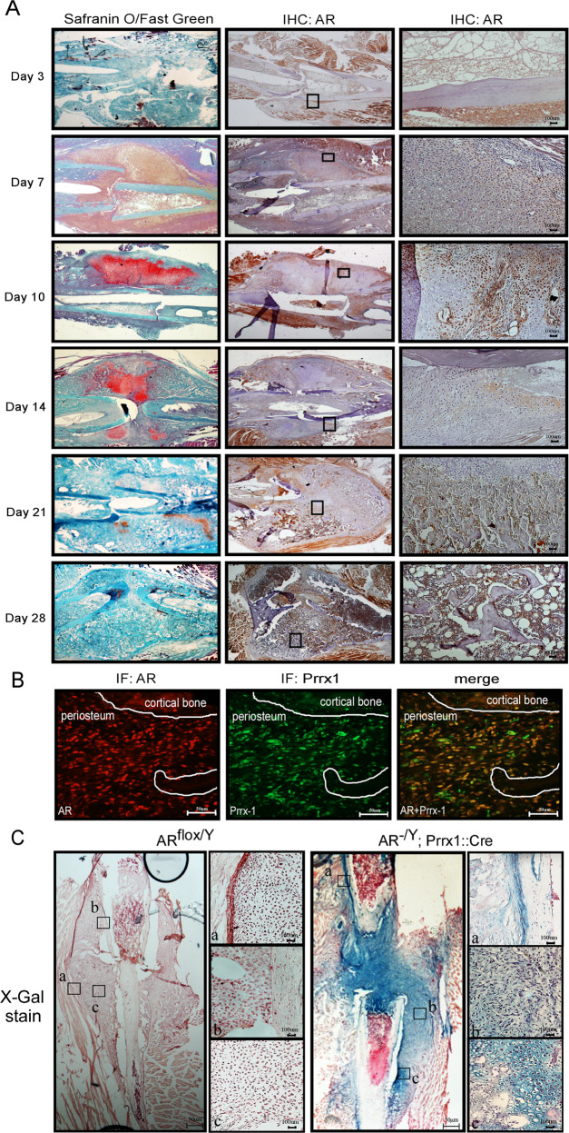
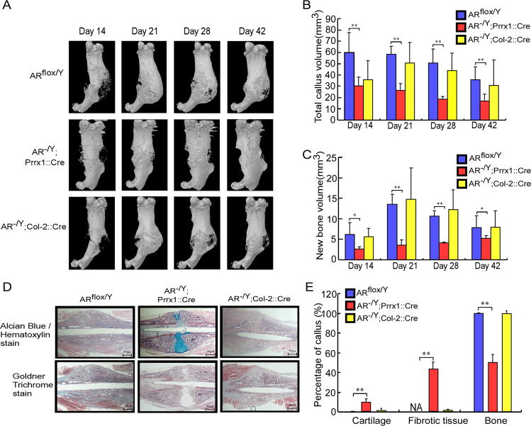
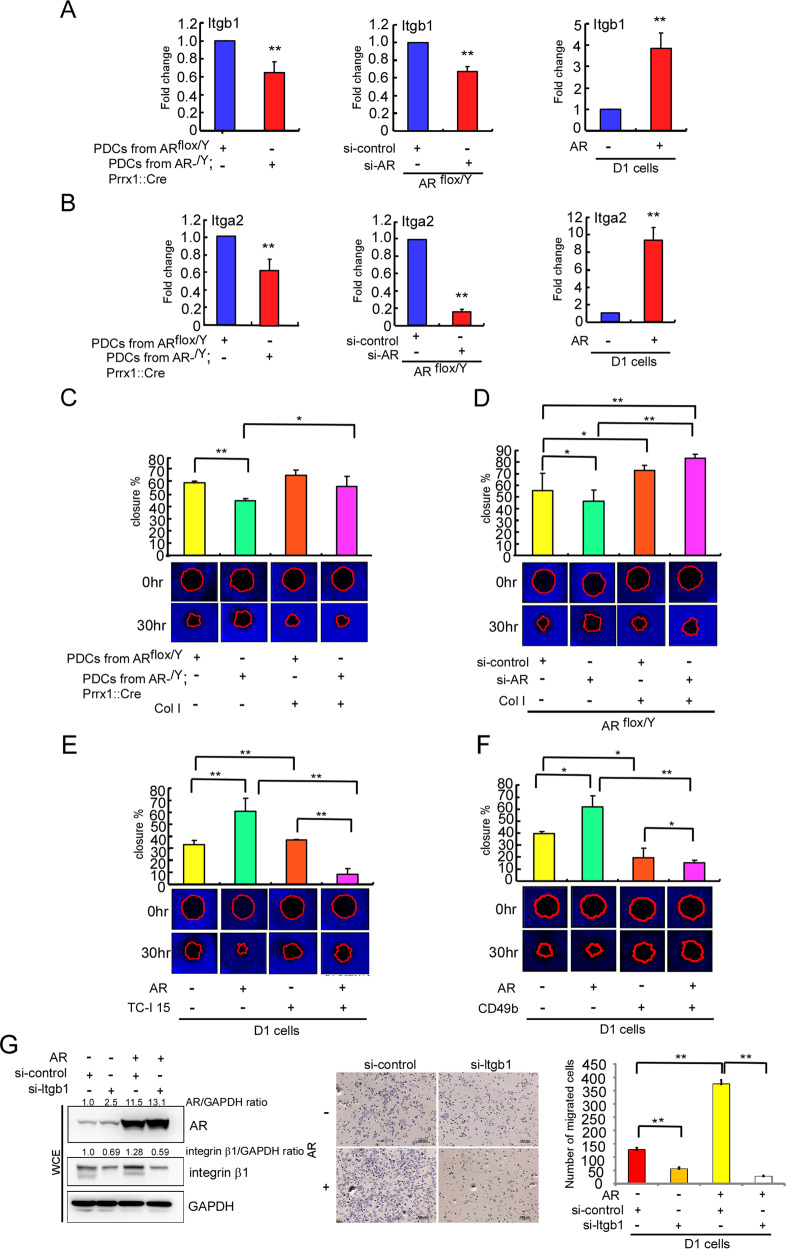
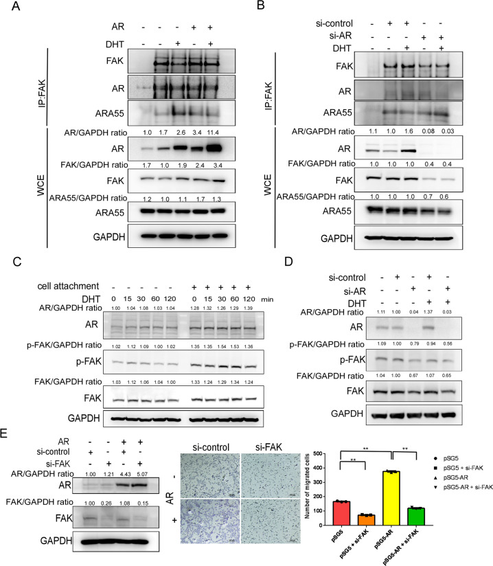
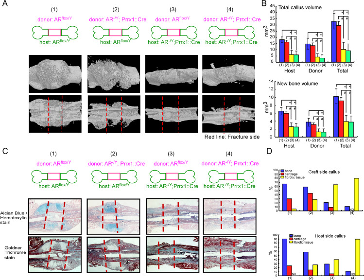
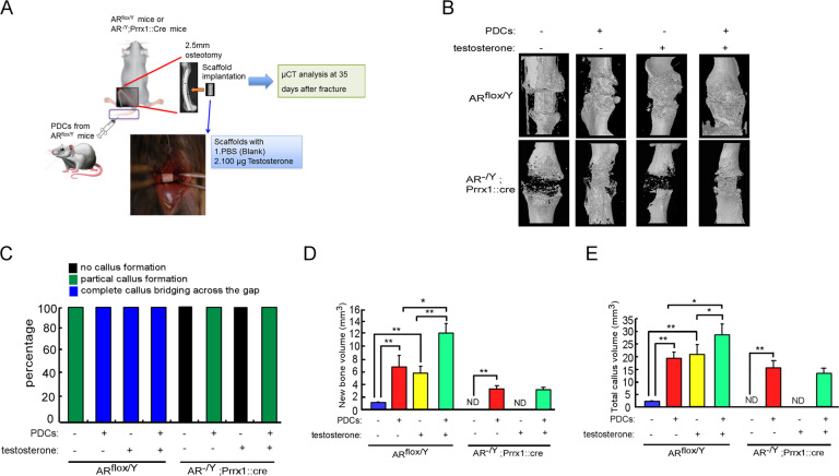
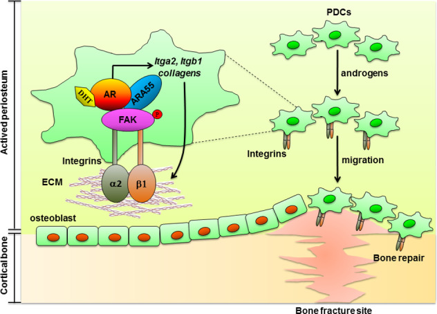
Low testosterone level is an independent predictor of osteoporotic fracture in elderly men as well as increased fracture risk in men undergoing androgen deprivation. Androgens and androgen receptor (AR) actions are essential for bone development and homeostasis but their linkage to fracture repair remains unclear. Here we found that AR is highly expressed in the periosteum cells and is co-localized with a mesenchymal progenitor cell marker, paired-related homeobox protein 1 (Prrx1), during bone fracture repair. Mice lacking the AR gene in the periosteum expressing Prrx1-cre (AR-/Y;Prrx1::Cre) but not in the chondrocytes (AR-/Y;Col-2::Cre) exhibits reduced callus size and new bone volume. Gene expression data analysis revealed that the expression of several collagens, integrins and cell adhesion molecules were downregulated in periosteum-derived progenitor cells (PDCs) from AR-/Y;Prrx1::Cre mice. Mechanistically, androgens-AR signaling activates the AR/ARA55/FAK complex and induces the collagen-integrin α2β1 gene expression that is required for promoting the AR-mediated PDCs migration. Using mouse cortical-defect and femoral graft transplantation models, we proved that elimination of AR in periosteum of host mice impairs fracture healing, regardless of AR existence of transplanted donor graft. While testosterone implanted scaffolds failed to complete callus bridging across the fracture gap in AR-/Y;Prrx1::Cre mice, cell-based transplantation using DPCs re-expressing AR could lead to rescue bone repair. In conclusion, targeting androgen/AR axis in the periosteum may provide a novel therapy approach to improve fracture healing.


Conflict of interest statement

The authors declare no competing interests.

Figures

Fig. 1
Fig. 1. The AR is expressed in the periosteum during the fracture healing process.
A Histological sections of post-fracture calluses at 3, 7, 10, 14, 21 and 28 days were analyzed by Safranin O/Fast Green staining and immunohistochemical analysis of AR expression. Scale bar = 100 µm. B Double-immunofluorescence staining revealed co-localization of the AR (red) and Prrx1 (green) in the periosteum of post-fracture calluses at 10 days. Scale bar = 50 µm. C X-gal–stained cells were observed in the post-fracture callus at 10 days in AR-/Y;Prrx1::Cre::Rosa26-LacZ mice (right panel); and ARflox/Y mice (left panel) were used as negative controls. Scale bar = 50 µm. a. periosteum; b. mesenchyme; c. chondrocytes. Scale bar = 100 µm.
Fig. 2
Fig. 2. Prrx1-specific AR knockout (AR-/Y;Prrx1::Cre) mice have decreased callus volume and new bone volume in the fracture callus during fracture repair.
A Representative images of micro-CT 3D reconstructions of the fracture site 14–42 days after fracture in ARflox/Y, AR-/Y;Prrx1::Cre and AR-/Y;Col-2::Cre mice. B, C Mineralized callus formation in ARflox/Y, AR-/Y;Prrx1::Cre and AR-/Y;Col-2::Cre mice, determined by quantitative analysis of total callus bone volume and new bone volume. D After micro-CT analysis, the cross-sections of 21-day fracture calluses were stained with Alcian Blue /Hematoxylin and Goldner Trichrome stain. Scale bar = 50 µm. E The percentages of cartilage, fibrotic tissue, and bone were quantified. Data are presented as mean ± SEM (n ≥ 3; *P < 0.05, **P < 0.001 compared with ARflox/Y mice, one-way ANOVA).
Fig. 3
Fig. 3. Collagens-integrins signaling is essential for AR to enhance the migration potential of PDCs in vitro.
Primary PDCs were isolated from ARflox/Y and AR-/Y;Prrx1::Cre mice. The AR in PDCs from ARflox/Y mice was knocked down using AR-targeted siRNA (si-AR). D1 cells were transfected with Flag (pcDNA3-flag) or Flag-tagged AR (pcDNA3-flag-AR). A, B Itgb1 and Itga2 mRNA levels were quantified by RT-qPCR. C, D Cells were cultured with or without the integrin ligand, Col I. E, F Cells were incubated with or without the integrin α2β1 inhibitor TC-I or integrin α2β1 blocking antibody CD49b. (C–F) Migration capacity was measured using the OrisTM Cell Migration Assay (CMA) kit. G D1 cells were transfected with pSG5 or AR (pSG5-AR). The integrin β1 in the transfected cells was knocked down using Itgb1-targeted siRNA (si-Itgb1). Cell migration assay was stained with Giemsa stain. AR, integrin β1 and GAPDH protein levels were then determined by western blot analysis. Experiments were conducted three times. Data were expressed as the mean ± SD. Statistical correlation of data was checked for significance by Student’s t test. *P < 0.05, **P < 0.001 was considered to indicate a statistically significance result.
Fig. 4
Fig. 4. AR interacts with FAK/ARA55 complexes and FAK is required for AR-mediated cell migration.
A, B AR-overexpressing D1 cells (pcDNA3-flag-AR transfection) and AR-knockdown D1 cells (si-AR) were treated with 10 nM DHT for 15 min. FAK, AR, ARA55 and β-tubulin protein levels were then determined by western blot analysis. Cell lysates were also immunoprecipitated (IP) with anti-AR or anti-FAK antibodies, after which immunoprecipitates were analyzed for interacting proteins (FAK, AR, ARA55) by western blotting. C D1 cells were exposed to 10 nM DHT for 15–120 min with or without the attachment. Phosphorylation of FAK at Tyr-397 (p-FAK PY397), total FAK and AR were assayed by western blot analysis. GAPDH was used as a loading control. D D1 cells were knocked down using si-AR and exposed to 10 nM DHT. Phosphorylation of FAK at Tyr-397 (p-FAK PY397), total FAK and AR were assayed by western blot analysis. GAPDH was used as a loading control. E D1 cells were transfected with pSG5 or AR (pSG5-AR). The FAK in the transfected cells was knocked down using FAK-targeted siRNA (si-FAK). Cell migration assay was stained with Giemsa stain. Experiments were conducted three times. Data were expressed as the mean ± SD. Statistical correlation of data was checked for significance by Student’s t test. *P < 0.05, **P < 0.001 was considered to indicate a statistically significance result. AR, FAK and GAPDH protein levels were then determined by western blot analysis.
Fig. 5
Fig. 5. Femoral bone graft transplantations showed that deletion of AR in Prrx1-cre expressing cells from host mice impairs the function of ARflox/Y donor graft.
ARflox/Y and AR-/Y;Prrx1::Cre host mice received transplantations of bone grafts from ARflox/Y or AR-/Y;Prrx1::Cre donor mice and samples were acquired on 14 days after the operation. A Representative 3D micro-CT images of femoral bone graft transplantation between ARflox/Y (WT, wildtype) and AR-/Y;Prrx1::Cre (ARKO) mice are shown: (1) WT graft to WT host, (2) KO graft to WT host, (3) WT graft to KO host, (4) KO graft to KO host. B Quantitative micro-CT analyses of total callus volume and new bone volume on host bone graft, donor bone graft and total bone segments are shown. Data are presented as mean ± SEM (n ≥ 5; *P < 0.001, #P < 0.001, one-way ANOVA). C Representative images of cross-sections of 14-day fracture calluses from four different groups stained with Alcian Blue/Hematoxylin and Goldner Trichrome stain. Scale bar = 50 µm. D Histomorphometric analyses of callus were performed on histologic sections prepared from four groups of transplantations. The percent area of bone (blue bar), cartilage (red bar) and fibrotic tissue (yellow bar) on the graft side (upper panel) and host side (lower panel) were quantified.
Fig. 6
Fig. 6. Combination of androgen therapy with the DPCs cell-based transplantation augments the repair of segmental bone defects.
ARflox/Y and AR-/Y;Prrx1::Cre host mice received transplantations of bone grafts from ARflox/Y or AR-/Y;Prrx1::Cre donor mice and samples were acquired on 14 days after the operation. A ARflox/Y and AR-/Y;Prrx1::Cre mice received segmental bone defects, and then were implanted with scaffold containing vehicle control or 100 μg testosterone. These host mice were also treated with PDCs from ARflox/Y donor mice by tail vein injection. B 3D micro-CT images of defect sites are shown at 35 days post operation. C The percentages of no callus formation, partial callus formation and complete callus bridging across the gap were quantified by X-ray image analysis. D, E The new bone volume and total callus volume were quantified by micro-CT analysis. Data are presented as mean ± SEM (n ≥ 3; *P < 0.05, **P < 0.001, one-way ANOVA). ND not detected.
Fig. 7
Fig. 7. Novel androgen-AR-targeted approaches that promote homing of PDCs to bone formation and repair.
The targeted androgens-AR and collagen-integrin α2β1 in PDCs can increase the formation of AR/ARA55/FAK complex that resulted promote the PDCs migration via activation of signaling mediated by ECM–integrin interactions, an effect that translates into increased periosteal bone formation and improved bone fracture repair. AR androgen receptor, ARA55 AR-associated protein 55, FAK focal adhesion kinase, ECM extracellular matrix, PDCs periosteum-derived progenitor cells.

References

    1. Loi F, Cordova LA, Pajarinen J, Lin TH, Yao Z, Goodman SB. Inflammation, fracture and bone repair. Bone. 2016;86:119–30. - PMC - PubMed
    1. Evans SF, Chang H, Knothe Tate ML. Elucidating multiscale periosteal mechanobiology: a key to unlocking the smart properties and regenerative capacity of the periosteum? Tissue Eng Part B Rev. 2013;19:147–59. - PMC - PubMed
    1. Murao H, Yamamoto K, Matsuda S, Akiyama H. Periosteal cells are a major source of soft callus in bone fracture. J Bone Min Metab. 2013;31:390–8. - PubMed
    1. Zhang X, Awad HA, O’Keefe RJ, Guldberg RE, Schwarz EM. A perspective: engineering periosteum for structural bone graft healing. Clin Orthop Relat Res. 2008;466:1777–87. - PMC - PubMed
    1. Colnot C. Skeletal cell fate decisions within periosteum and bone marrow during bone regeneration. J Bone Miner Res. 2009;24:274–82. - PMC - PubMed

Publication types