. 2022 Feb 8;13(2):126.
doi: 10.1038/s41419-022-04578-2.

LncRNA BC200/miR-150-5p/MYB positive feedback loop promotes the malignant proliferation of myelodysplastic syndrome

Affiliations

LncRNA BC200/miR-150-5p/MYB positive feedback loop promotes the malignant proliferation of myelodysplastic syndrome

Zhaoping Liu et al. Cell Death Dis. .

Abstract

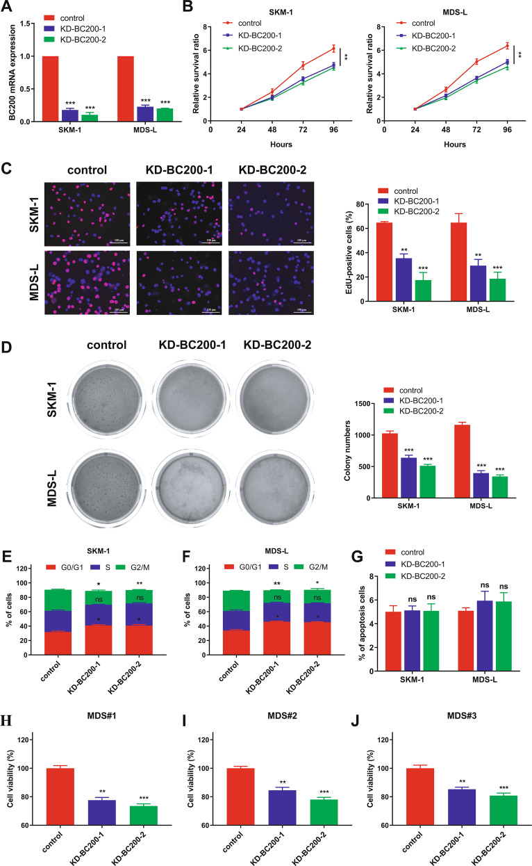
Myelodysplastic syndrome (MDS) is a group of heterogeneous hematologic malignancies with a risk of transformation to acute myeloid leukemia. Understanding the molecular mechanisms of the specific roles of long noncoding RNAs (lncRNAs) in MDS would create novel ways to identify diagnostic and therapeutic targets. The lncRNA BC200 is upregulated and acts as an oncogene in various cancers; however, its expression, clinical significance, and roles in MDS remain unclear. Here, we found that BC200 was highly expressed in MDS patients compared with normal individuals. Knockdown of BC200 inhibited MDS cell proliferation, colony formation, and cell cycle progression in vitro and suppressed the growth and invasiveness of MDS cells in vivo. Mechanistic investigations revealed that BC200 functioned as a miRNA sponge to positively regulate the expression of MYB through sponging miR-150-5p and subsequently promoted malignant proliferation of MDS cells. Conversely, we found that BC200 was a direct transcriptional target of MYB, and knockdown of MYB abolished the oncogenic effect of BC200/miR-150-5p. Taken together, our results revealed that the BC200/miR-150-5p/MYB positive feedback loop promoted the proliferation of MDS cells and is expected to be a potential biomarker and therapeutic target in MDS.


Conflict of interest statement

The authors declare no competing interests.

Figures

Fig. 1
Fig. 1. Knockdown of BC200 inhibits the proliferation of MDS cells.
A In SKM-1 and MDS-L cells, the transcriptional level of BC200 was downregulated by two shRNAs. B–D MDS cells proliferation was detected by CCK-8, EdU incorporation (Scale bar, 100 μm) and colony formation assays. It revealed that BC200 knockdown significantly suppressed SKM-1 and MDS-L cell proliferation. E, F Knockdown of BC200 led to G0/G1 arrest in both SKM-1 and MDS-L cells. G Apoptosis assay showed that the percentage of apoptotic MDS cells was not affected by BC200 knockdown. H–J Human primary BMMCs were treated with BC200 siRNAs for 72 h, and cell proliferation was analyzed by the CCK-8 assay. *p < 0.05, **p < 0.01, ***p < 0.001, ns, not significant.
Fig. 2
Fig. 2. BC200 acts as a molecular sponge for miR-150-5p in MDS cells.
A RNA FISH assays for BC200 in SKM-1 cells. Stain the nucleus with DAPI. Scale bar, 25 μm. B BC200, U6, and ACTB expression in the RNA extracted from the cytoplasm and nucleus of MDS cells by qRT-PCR. C The ceRNA regulatory network of BC200 and three miRNAs containing the binding site of BC200. D Transcriptional level of miR-150-5p was detected in sh-BC200 MDS cells. E Upper: complementary sequence between miR-150-5p and BC200-wt. The putative binding sites of miR-150-5p in BC200-mt. Lower: Luciferase activity was measured in HEK293T cells cotransfected with miR-150-5p mimics and wt or mt BC200 vector. F RIP assays of the enrichment of Ago2 on BC200 and miR-150-5p relative to IgG in MDS-L cells. The relative expression levels of BC200 and miR-150-5p were detected by qRT-PCR. G Detection of BC200 or miR-150-5p by using qRT-PCR in the sample pulled down by biotinylated BC200 and Random probe. Input was used for normalization. H The correlation among BC200, miR-150-5p, and Ago2 was determined by using Ago2 antibody to detect cell lysates utilizing the sample pulled down by biotinylated BC200 and Random probe. I CCK-8 assay rescue experiment showed that cell proliferation reduced by si-BC200 could be increased by miR-150-5p inhibitor in MDS cells. J CCK-8 assay rescue experiment showed that cell proliferation stimulated by overexpression of BC200 could be suppressed by miR-150-5p mimics in MDS cells. *p < 0.05, **p < 0.01, ***p < 0.001, ns, not significant.
Fig. 3
Fig. 3. MYB is a direct target of miR-150-5p in MDS cells.
A MYB was the top candidate target containing the complementary site for the seed region of miR-150-5p in the TargetScan database. B, C SKM-1 and MDS-L cells were transfected with miR-150-5p mimics or inhibitors for 48 h, and MYB mRNA levels were detected by qRT-PCR. D, E MYB protein expression in both SKM-1 and MDS-L cells transfected with miR-150-5p mimics or inhibitor for 48 h was detected by western blotting. F Upper: putative MYB binding sites on the promoter region of miR-150-5p and their corresponding mutant binding sites. Lower: luciferase activity in HEK293T cells cotransfected with miR-150-5p or control mimics and luciferase reporters containing the wt or mt MYB 3'-UTR. G CCK-8 assay detected the effect of MYB overexpression and miR-150-5p mimics on SKM-1 and MDS-L cell proliferation activity after 72 h. H CCK-8 assay detected the effect of MYB knockdown and miR-150-5p inhibitor on SKM-1 and MDS-L cell proliferation activity after 72 h. *p < 0.05, **p < 0.01, ***p < 0.001, ns, not significant.
Fig. 4
Fig. 4. BC200 promotes the proliferation of MDS cells via the miR-150-5p/MYB axis.
A The mRNA levels of MYB were measured in sh-BC200 MDS cells. B The protein expression levels of MYB were measured in MDS cells transfected with two different BC200 shRNAs. C, D qRT-PCR and western blotting were used to measure the MYB mRNA and protein levels in SKM-1 and MDS-L cells transfected with BC200 plasmid (ov-BC200) for 48 h. E, F Western blot analysis of MYB in MDS cells cotransfected with miR-150-5p mimics and BC200 siRNA or miR-150-5p inhibitors and BC200 plasmid for 48 h. G CCK-8 assay indicated the effect of BC200 overexpression and MYB suppression on SKM-1 and MDS-L cell proliferation activity after 72 h. H CCK-8 assay indicated the effect of BC200 knockdown and MYB upregulation on SKM-1 and MDS-L cell proliferation activity after 72 h. *p < 0.05, **p < 0.01, ***p < 0.001, ns, not significant.
Fig. 5
Fig. 5. MYB transcriptionally regulates BC200 expression in MDS cells.
A HEK293T cells were cotransfected with GV238-BC200 and MYB siRNAs for 48 h. B The MYB binding sitein BC200 predicted by JASPAR matrix models. C Upper: putative MYB binding sites BC200 promoter region and design of the indicated primers. Lower: ChIP assays of the enrichment of MYB on the BC200 promoter relative to control IgG in MDS-L cells. D The expression level of BC200 was detected in MDS cells transfected with two different MYB siRNAs. E, F SKM-1 and MDS-L cells were treated with the indicated siRNAs or plasmids for 72 h. The cell proliferative was measured by CCK-8 assay. *p < 0.05, **p < 0.01, ***p < 0.001, ns, not significant.
Fig. 6
Fig. 6. Knockdown of BC200 inhibits MDS growth in vivo.
A SKM-1 cells treated with different BC200 shRNAs (n = 4) or control shRNA (n = 4) were inoculated subcutaneously into NCG mice. Tumor growth curves showed that sh-BC200 group led to tumor growth restriction in mice. Scale bar: 1 cm. B Tumor growth curves showed that sh-BC200 group suppressed tumor growth compared with sh-control group. C The subcutaneous tumors were harvested and weighed on the 26th day after implantation. Data are shown as mean ± SEM. D The NCG mice grew tumors on the fourteenth day after implantation and began to be weighed. E Ki-67 immunostaining of xenograft tissues collected from the BC200-knockdown group and control group. Scale bar: 50 μm. F–H qRT-PCR was performed on xenograft tissues and was subjected to measure BC200, MYB, and miR-150-5p expression. I Plot of CD123 vs CD34 expression of BM cells of MDS or normal mice being analyzed. J White blood cells (WBCs), red blood cells (RBCs), hemoglobin (HGB), hematocrit (HCT), and platelets (PLTs) counts of MDS or normal mice. *p < 0.05, **p < 0.01, ***p < 0.001, ****p < 0.0001.
Fig. 7
Fig. 7. Clinical significance of the BC200/miR-150-5p/MYB loop based on bioinformatics analysis.
A–C Analysis of a GEO dataset (GSE114869) indicated that BC200 and MYB expression was markedly higher in MDS samples than in normal samples. Conversely, miR-150-5p was significantly lower in MDS samples. D–F The expression of BC200, miR-150-5p mRNA, and MYB was analyzed by qRT-PCR in 79 MDS samples and 44 normal samples. G–I Correlation analysis of BC200 and miR-150-5p, MYB and miR-150-5p, and BC200 and MYB expression levels in 75 MDS samples. ****p < 0.0001.
Fig. 8
Fig. 8. Schematic diagram of the BC200/miR-150-5p/MYB positive feedback loop in promoting the proliferation of MDS cells.
BC200 acts as a miRNA sponge and positively regulates the expression of MYB through sponging miR-150-5p. Reciprocally, MYB can transcriptionally regulate the expression of BC200, and the overexpression of MYB enhanced the oncogenic effect of BC200/miR-150-5p. Therefore, BC200/miR-150-5p/MYB positive feedback loop is involved in promoting the malignant proliferation of MDS cells.

References

    1. Steensma DP. Myelodysplastic syndromes: diagnosis and treatment. Mayo Clin Proc. 2015;90:969–83.. - PubMed
    1. Kennedy JA, Ebert BL. Clinical implications of genetic mutations in myelodysplastic syndrome. J Clin Oncol. 2017;35:968–74.. - PMC - PubMed
    1. Elnahass Y, Youssif L. Cytogenetic features in primary myelodysplastic syndrome Egyptian patients. J Adv Res. 2018;10:77–83. - PMC - PubMed
    1. He Q, Zheng Q, Xu F, Shi W, Guo J, Zhang Z, et al. IGF‑IR promotes clonal cell proliferation in myelodysplastic syndromes via inhibition of the MAPK pathway. Oncol Rep. 2020;44:1094–104.. - PMC - PubMed
    1. Kam AYF, Piryani SO, McCall CM, Park HS, Rizzieri DA, Doan PL. Targeting high mobility group box-1 (HMGB1) promotes cell death in myelodysplastic syndrome. Clin Cancer Res. 2019;25:4155–67.. - PMC - PubMed

Publication types