. 2022 Feb 8;13(2):124.
doi: 10.1038/s41419-022-04550-0.

Pharmacological inhibition of Ref-1 enhances the therapeutic sensitivity of papillary thyroid carcinoma to vemurafenib

Affiliations

Pharmacological inhibition of Ref-1 enhances the therapeutic sensitivity of papillary thyroid carcinoma to vemurafenib

Linfei Hu et al. Cell Death Dis. .

Abstract

The use of the BRAF inhibitor vemurafenib exhibits drug resistance in the treatment of thyroid cancer (TC), and finding more effective multitarget combination therapies may be an important solution. In the present study, we found strong correlations between Ref-1 high expression and BRAF mutation, lymph node metastasis, and TNM stage. The oxidative stress environment induced by structural activation of BRAF upregulates the expression of Ref-1, which caused intrinsic resistance of PTC to vemurafenib. Combination inhibition of the Ref-1 redox function and BRAF could enhance the antitumor effects of vemurafenib, which was achieved by blocking the action of Ref-1 on BRAF proteins. Furthermore, combination treatment could cause an overload of autophagic flux via excessive AMPK protein activation, causing cell senescence and cell death in vitro. And combined administration of Ref-1 and vemurafenib in vivo suppressed PTC cell growth and metastasis in a cell-based lung metastatic tumor model and xenogeneic subcutaneous tumor model. Collectively, our study provides evidence that Ref-1 upregulation via constitutive activation of BRAF in PTC contributes to intrinsic resistance to vemurafenib. Combined treatment with a Ref-1 redox inhibitor and a BRAF inhibitor could make PTC more sensitive to vemurafenib and enhance the antitumor effects of vemurafenib by further inhibiting the MAPK pathway and activating the excessive autophagy and related senescence process.


Conflict of interest statement

The authors declare no competing interests.

Figures

Fig. 1
Fig. 1. Ref-1 expression was upregulated in PTC patient samples.
A Amplification alteration of the APEX1 gene in thyroid cancer and normal tissue samples from The Cancer Genome Atlas (TCGA) database. B Real-time PCR detection of the mRNA expression level of Ref-1 in tumor and normal tissue samples from 16 patients. C Western blot detection of the protein expression level of Ref-1 in tumor and normal tissue specimens from 8 patients. D Representative immunohistochemical staining for Ref-1 in PTC specimens. E Percentages of samples with different BRAF mutation statuses and specific Ref-1 expression levels among 178 PTC cases. F RFS analysis of groups based on the high and low expression of Ref-1 among 178 PTC cases. *P < 0.05, **P < 0.01.
Fig. 2
Fig. 2. Resistance to vemurafenib in PTC was related to upregulated Ref-1 expression.
A Analysis of data from the TCGA database for Ref-1 mRNA expression levels in BRAFwt and BRAFmut PTC (upper) and melanoma (bottom) cases. B Correlation analysis of Ref-1 and MAPK pathway targeting genes expression in PTC (upper) and melanoma (bottom) cases based on the TCGA database. C Analysis of data from the TCGA database for NOX4 mRNA expression levels in BRAFwt and BRAFmut PTC (upper) and melanoma (bottom) cases. D Correlation analysis of Ref-1 and NOX4 mRNA expression levels in PTC (upper) and melanoma (bottom) cases based on the TCGA database. E Vemurafenib IC50 detection by a CCK-8 assay in BCPAP, K-1, and A375 cells. F Cell proliferation ability detection by a CCK-8 assay after vemurafenib (10 μM) and E3330 (different concentration gradient) pretreatment in BCPAP and K-1 cells. G Vemurafenib IC50 detection after E3330 pretreatment by a CCK-8 assay in BCPAP and K-1 cells. *P < 0.05, **P < 0.01. ***P < 0.001.
Fig. 3
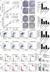
Fig. 3. E3330 enhanced the antiproliferative capacity of vemurafenib in BCPAP and K-1 cells.
A, B Cell viability detection by a CCK-8 assay was used to quantify cell proliferation in BCPAP and K-1 cell lines. C, D Cell proliferation detection by colony-formation assay in BCPAP and K-1 cell lines. E, F Cell division detection by BrdU assay in BCPAP and K-1 cell lines. G, H The cell cycle distribution detection by flow cytometric assay in BCPAP and K-1 cell lines. BCPAP and K-1 cell lines were pretreated with DMSO, vemurafenib (10 μM), E3330 (50 μM), and combination (10 μM vemurafenib + 50 μM E3330), respectively. *P < 0.05, **P < 0.01, ***P < 0.001.
Fig. 4
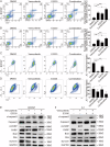
Fig. 4. E3330 enhanced the proapoptotic capacity of vemurafenib in BCPAP and K-1 cells.
A, B The cell apoptosis rate measured by flow cytometric analysis in BCPAP and K-1 cell lines. C, D The mitochondrial membrane potential evaluation by detecting JC-1 content in BCPAP and K-1 cell lines. E, F Mitochondrial apoptosis pathway-associated protein expression detection by western blot assay in BCPAP and K-1 cell lines. BCPAP and K-1 cell lines were pretreated with DMSO, vemurafenib (10 μM), E3330 (50 μM), and combination (10 μM vemurafenib + 50 μM E3330), respectively. *P < 0.05, **P < 0.01, ***P < 0.001.
Fig. 5
Fig. 5. Ref-1 promoted the maintenance of mutant BRAF function.
A, B Western blot detection of MAPK pathway activation in DMSO, vemurafenib (10 μM), E3330 (50 μM), and combination (10 μM vemurafenib + 50 μM E3330) groups. C The mode of action of Ref-1 and BRAF determination by a Co-IP assay using TPC-1 cells with a BRAFV600E Tet-on system. D MAPK pathway activation detection by western blot assay after small interfering RNA targeting of Ref-1 with vemurafenib treatment.
Fig. 6
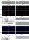
Fig. 6. Combined Ref-1 redox inhibitor and vemurafenib treatment induced autophagic flow overload and caused senescence in BCPAP and K-1 cells.
A Representative electron micrographs of BCPAP and K-1 cell lines. The green arrows indicate lysosome and the red arrows indicate autophagosome. B Immunofluorescence detection of the autophagic substrates LC3B and p62 in BCPAP and K-1 cell lines. C Western blot detection of the autophagic substrates LC3B and p62 in BCPAP and K-1 cell lines. D Cellular senescence detection by β-galactosidase staining in BCPAP and K-1 cell lines. E Immunofluorescence detection of γH2AX in BCPAP and K-1 cell lines. F Western blot detection of the autophagic substrates LC3B and p62 in BCPAP and K-1 cell lines. G Cellular senescence detection by β-galactosidase staining in BCPAP and K-1 cell lines. BCPAP and K-1 cell lines in AE were pretreated with DMSO, vemurafenib (10 μM), E3330 (50 μM), and combination (10 μM vemurafenib + 50 μM E3330), respectively. BCPAP and K-1 cell lines in F, G were pretreated with DMSO, combination (10 μM vemurafenib + 50 μM E3330), HCQ, and combination + HCQ, respectively.
Fig. 7
Fig. 7. Vemurafenib combined with E3330 treatment offered a relatively promising therapeutic strategy in vivo.
A Representative images of dissected mouse subcutaneous tumors after HMC, vemurafenib, E3330, or combination (vemurafenib + E3330) treatment for 21 days. B The weights of mouse subcutaneous tumors after different treatments for 21 days. C Change in tumor volume after HMC, vemurafenib, E3330, or combination (vemurafenib + E3330) treatment for 21 days. D The body weight changes of mice measured every 3 days after different treatments. E Representative H&E staining of tumors, livers, and kidneys after different treatments. F Representative immunohistochemical staining for p-ERK, Ki-67, cl-caspase3, and γH2AX after different treatments. G Representative images of mouse metastatic tumors after HMC, vemurafenib, E3330, or combination (vemurafenib + E3330) treatment for 21 days. H Representative H&E staining of metastatic tumors in the lungs and livers. I Representative immunohistochemical staining for Ki-67, Vimentin, LC3B, and p62 after different treatments. **P < 0.01, ***P < 0.001.

Similar articles

Cited by

References

    1. Proietti I, Skroza N, Michelini S, Mambrin A, Balduzzi V, Bernardini N, et al. BRAF inhibitors: molecular targeting and immunomodulatory actions. Cancers. 2020;12:1823. - PMC - PubMed
    1. Patel H, Yacoub N, Mishra R, White A, Long Y, Alanazi S, et al. Current advances in the treatment of BRAF-mutant melanoma. Cancers. 2020;12:482. - PMC - PubMed
    1. Fujimura T, Fujisawa Y, Kambayashi Y, Aiba S. Significance of BRAF kinase inhibitors for melanoma treatment: from bench to bedside. Cancers. 2019;11:1342. - PMC - PubMed
    1. Dadu R, Shah K, Busaidy NL, Waguespack SG, Habra MA, Ying AK, et al. Efficacy and tolerability of vemurafenib in patients with BRAF(V600E)-positive papillary thyroid cancer: MD Anderson Cancer Center off label experience. J Clin Endocrinol Metab. 2015;100:E77–81. - PMC - PubMed
    1. Kim KB, Cabanillas ME, Lazar AJ, Williams MD, Sanders DL, Ilagan JL, et al. Clinical responses to vemurafenib in patients with metastatic papillary thyroid cancer harboring BRAF(V600E) mutation. Thyroid. 2013;23:1277–83. - PMC - PubMed

Publication types

MeSH terms

Substances