. 2022 Feb 8;13(1):750.
doi: 10.1038/s41467-022-28358-w.

In vivo mitochondrial base editing via adeno-associated viral delivery to mouse post-mitotic tissue

Affiliations

In vivo mitochondrial base editing via adeno-associated viral delivery to mouse post-mitotic tissue

Pedro Silva-Pinheiro et al. Nat Commun. .

Abstract

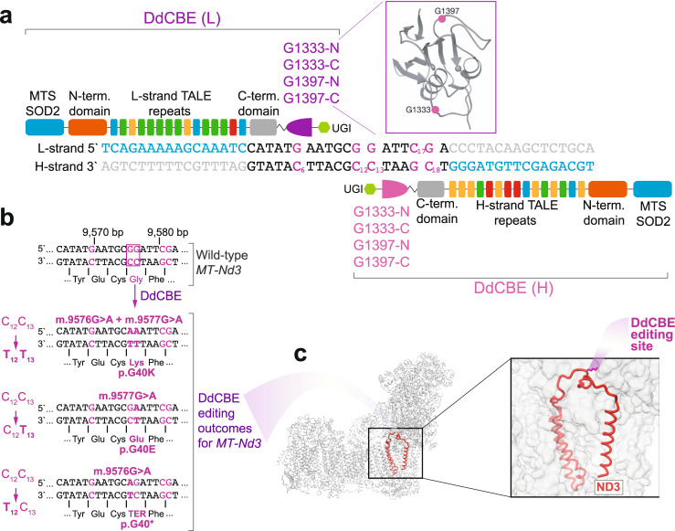
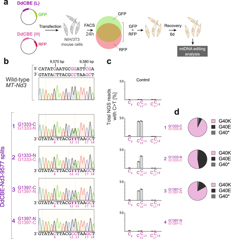
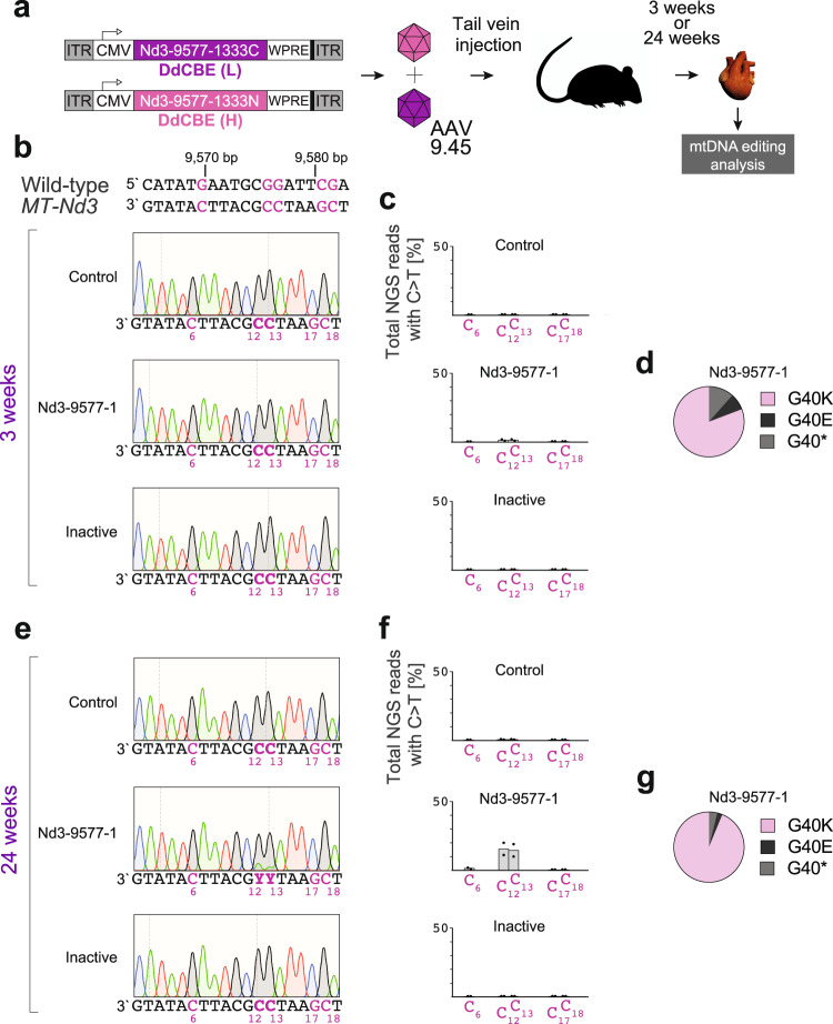
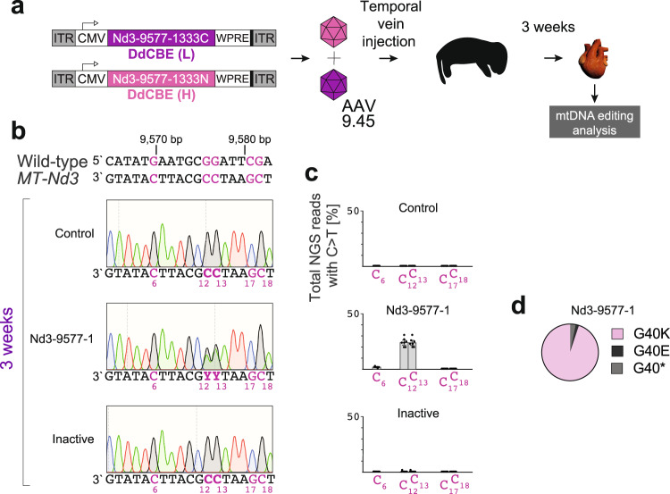
Mitochondria host key metabolic processes vital for cellular energy provision and are central to cell fate decisions. They are subjected to unique genetic control by both nuclear DNA and their own multi-copy genome - mitochondrial DNA (mtDNA). Mutations in mtDNA often lead to clinically heterogeneous, maternally inherited diseases that display different organ-specific presentation at any stage of life. For a long time, genetic manipulation of mammalian mtDNA has posed a major challenge, impeding our ability to understand the basic mitochondrial biology and mechanisms underpinning mitochondrial disease. However, an important new tool for mtDNA mutagenesis has emerged recently, namely double-stranded DNA deaminase (DddA)-derived cytosine base editor (DdCBE). Here, we test this emerging tool for in vivo use, by delivering DdCBEs into mouse heart using adeno-associated virus (AAV) vectors and show that it can install desired mtDNA edits in adult and neonatal mice. This work provides proof-of-concept for use of DdCBEs to mutagenize mtDNA in vivo in post-mitotic tissues and provides crucial insights into potential translation to human somatic gene correction therapies to treat primary mitochondrial disease phenotypes.


Conflict of interest statement

M.M. is a co-founder, shareholder and member of the Scientific Advisory Board of Pretzel Therapeutics, Inc. P.S.-P., P.A.N., and C.D.M. provide consultancy services for Pretzel Therapeutics, Inc. The remaining authors declare no competing interests.

Figures

Fig. 1
Fig. 1. Design of DdCBEs and mutagenesis site.
a The architecture of DdCBE monomers targeting m.9576 G (C12) and m.9577 G (C13). The DNA specificity is provided by TALE domains. In each experiment, different DddAtox splits are used (G1397 or G1333, purple) to achieve editing of “TC” sites. MTS SOD2 mitochondrial targeting sequence from superoxide dismutase 2, UGI uracil glycosylase inhibitor, L-strand or (L) light mtDNA strand, H-strand or (H) heavy mtDNA strand. b The details and possible outcomes of m.9576 G (C12) and m.9577 G (C13) editing. The purple box indicates the desired editing sites; other potential editing sites are indicated in purple. c The structural model of mouse complex I with indicated MT-ND3 subunit (red). The inset shows the location of MT-ND3 p.G40K mutation on the evolutionary conserved MT-ND3 loop.
Fig. 2
Fig. 2. Mitochondrial DNA editing in cultured mouse cells.
a Schematic of the general workflow for in vitro experiments that involve transient transfection of cultured mouse NIH/3T3 cells with plasmids co-expressing DdCBE monomers and fluorescent marker proteins, FACS-based selection of cells expressing both monomers and evaluation of mtDNA from DdCBE-treated cells. b Editing of mouse MT-Nd3 with different DdCBE splits in cells 7 days post-transfection, analyzed by Sanger sequencing. Potential editing sites are indicated in purple. The C:G > T:A deamination leads to m.9576 G > A (C12 > T12) and m.9577 G > A (C13 > T13) mutations, which translate to p.G40K change in MT-ND3. c The NGS analysis of the editing region in cells treated with different DdCBE splits. Bars represent the mean (n = 2). Source data are provided as a Source Data file. The mutagenesis frequency for the catalytically inactive versions is provided in the Source Data file. d The distribution of NGS reads containing m.9576 G (C12) or m.9577 G (C13) edits. The G40K reads contain both m.9576 G > A (C12 > T12) and m.9577 G > A (C13 > T13) mutations, G40E reads contain only the m.9577 G > A (C13 > T13) mutation, while G40* reads contain only the m.9576 G > A (C12 > T12) mutation. Source data are provided as a Source Data file.
Fig. 3
Fig. 3. Mitochondrial DNA editing in adult mouse hearts.
a Scheme of in vivo experiments with adult mice. The DdCBE-Nd3-9577-1 monomers (see Fig. 2), and their catalytically inactive versions, were encoded in separate AAV genomes, encapsidated in AAV9.45 then simultaneously administered by tail-vein (TV) injection at 1 × 1012 vg/mouse of each monomer. Animals were sacrificed either 3 or 24-weeks post-injection and their cardiac tissue was examined for mtDNA editing. b, e Editing of mouse MT-Nd3 with DdCBE in mouse heart at 3-weeks (b) or 24-weeks (e) post-injection, analyzed by Sanger sequencing. Potential editing sites are indicated in purple. c, f The NGS analysis of the DdCBE editing within the targeted region in adult mouse hearts at 3 weeks (c) or 24 weeks (f) after injection. Bars represent the mean (n = 2). Source data are provided as a Source Data file. d, g The distribution of NGS reads containing m.9576 G (C12) or m.9577 G (C13) edits at 3 weeks (d) or 24 weeks (g) after injection. The G40K reads contain both m.9576 G > A (C12 > T12) and m.9577 G > A (C13 > T13) mutations, G40E reads contain only the m.9577 G > A (C13 > T13) mutation, while G40* reads contain only the m.9576 G > A (C12 > T12) mutation. Source data are provided as a Source Data file.
Fig. 4
Fig. 4. Mitochondrial DNA editing in neonatal mouse hearts.
a Scheme of in vivo experiments with neonatal mice. The DdCBE-Nd3-9577-1 monomers (see Fig. 2), and their catalytically inactive versions, were encoded in separate AAV genomes, encapsidated in AAV9.45 then simultaneously administered by temporal vein injection at 1 × 1012 vg/mouse of each monomer. Animals were sacrificed 3-weeks post-injection and their cardiac tissue was examined for mtDNA editing. b Editing of mouse MT-Nd3 with DdCBE in neonatal mouse heart at 3-weeks post-injection, analyzed by Sanger sequencing. Potential editing sites are indicated in purple. c The NGS analysis of the DdCBE editing within the targeted region in neonatal mouse hearts. Bars represent the mean and error bars represent ±SEM (n = 7). Source data are provided as a Source Data file. d The distribution of NGS reads containing m.9576 G (C12) or m.9577 G (C13) edits in neonatal hearts at 3-weeks post-injection. The G40K reads contain both m.9576 G > A (C12 > T12) and m.9577 G > A (C13 > T13) mutations, G40E reads contain only the m.9577 G > A (C13 > T13) mutation, while G40* reads contain only the m.9576 G > A (C12 > T12) mutation. Source data are provided as a Source Data file.

References

    1. Vafai SB, Mootha VK. Mitochondrial disorders as windows into an ancient organelle. Nature. 2012;491:374–383. doi: 10.1038/nature11707. - DOI - PubMed
    1. Suomalainen A, Battersby BJ. Mitochondrial diseases: the contribution of organelle stress responses to pathology. Nat. Rev. Mol. Cell Biol. 2018;19:77–92. doi: 10.1038/nrm.2017.66. - DOI - PubMed
    1. Rath S, et al. MitoCarta3.0: an updated mitochondrial proteome now with sub-organelle localization and pathway annotations. Nucleic Acids Res. 2021;49:D1541–D1547. doi: 10.1093/nar/gkaa1011. - DOI - PMC - PubMed
    1. Gorman GS, et al. Prevalence of nuclear and mitochondrial DNA mutations related to adult mitochondrial disease. Ann. Neurol. 2015;77:753–759. doi: 10.1002/ana.24362. - DOI - PMC - PubMed
    1. Russell OM, Gorman GS, Lightowlers RN, Turnbull DM. Mitochondrial diseases: hope for the future. Cell. 2020;181:168–188. doi: 10.1016/j.cell.2020.02.051. - DOI - PubMed

Publication types

Substances