. 2022 Feb 8;13(1):749.
doi: 10.1038/s41467-022-28427-0.

Tousled-like kinase 2 targets ASF1 histone chaperones through client mimicry

Affiliations

Tousled-like kinase 2 targets ASF1 histone chaperones through client mimicry

Bertrand Simon et al. Nat Commun. .

Abstract

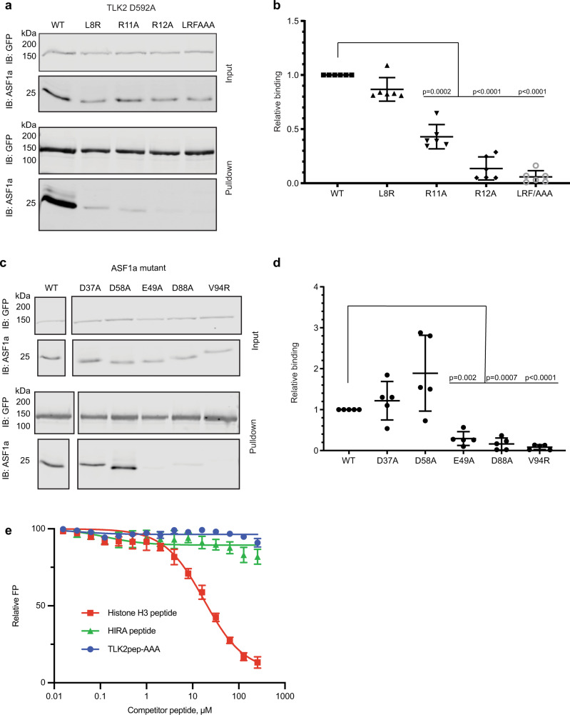
Tousled-like kinases (TLKs) are nuclear serine-threonine kinases essential for genome maintenance and proper cell division in animals and plants. A major function of TLKs is to phosphorylate the histone chaperone proteins ASF1a and ASF1b to facilitate DNA replication-coupled nucleosome assembly, but how TLKs selectively target these critical substrates is unknown. Here, we show that TLK2 selectivity towards ASF1 substrates is achieved in two ways. First, the TLK2 catalytic domain recognizes consensus phosphorylation site motifs in the ASF1 C-terminal tail. Second, a short sequence at the TLK2 N-terminus docks onto the ASF1a globular N-terminal domain in a manner that mimics its histone H3 client. Disrupting either catalytic or non-catalytic interactions through mutagenesis hampers ASF1 phosphorylation by TLK2 and cell growth. Our results suggest that the stringent selectivity of TLKs for ASF1 is enforced by an unusual interaction mode involving mutual recognition of a short sequence motifs by both kinase and substrate.


Conflict of interest statement

The authors declare no competing interests.

Figures

Fig. 1
Fig. 1. Optimal TLK2 phosphorylation of the C-terminal tails of ASF1 proteins is driven by both phosphorylation site specificity and the NT domain of ASF1.
a Heat map depicting amino acid selectivity at positions surrounding a Ser/Thr phosphorylation site determined from arrayed PSPL analysis of TLK2 ΔN178. The heat map shows normalized, log2 transformed PSPL spot intensities averaged across two separate experiments. Numerical values are provided in Supplementary Table 1. b Sequence logos showing positively selected residues were generated from the data shown in a using Seq2Logo. c Sequences of previously reported phosphorylation sites on human ASF1a and ASF1b. d Relative rates of TLK2 ΔN178 phosphorylation of a synthetic consensus peptide substrate (top) and Ala substitution variants (bars show mean ± SD, n = 4 independent experiments). e Representative assay showing TLK2 ΔN178 phosphorylation of purified GST-tagged ASF1b-CT (residues 155-202) and ASF1b-CT mutants was assessed in kinase assays in vitro. A kinase inactive TLK2-D592A mutant was included as a control. f Quantified results from three independent replicates of the experiment shown in panel e (mean ± SD) (right). g Electrostatic surface potential of the TLK2 and PKA catalytic domains (PDB 5O0Y and 1ATP) with overlayed peptide fragment from the PKA-PKI complex (PDB 1ATP) generated using Pymol. h Phosphorylation of purified GST-tagged FL ASF1b (FL), the N-terminal domain (NT, residues 1–155) or the C-terminal tail (CT, residues 155–202) by purified FL TLK2 (WT or kinase inactive D592A mutant) was assessed using in vitro kinase assays. Results from one representative experiment of two are depicted, along with a cartoon depicting ASF1b domain organization and major sites of phosphorylation. Source data are provided as a Source Data file.
Fig. 2
Fig. 2. The non-catalytic N-terminal region of TLK2 interacts with ASF1a.
a, b GFP, GFP-TLK2, or kinase-inactive GFP-TLK2 (D592A) was co-expressed with FLAG-tagged ASF1a in HEK293T cells, cells were lysed and GFP proteins were precipitated with anti-GFP nanobodies coupled to Sepharose beads. Precipitated GFP proteins and co-precipitating FLAG-ASF1a were fractionated by SDS-PAGE and detected by immunoblotting. A representative experiment is shown in a. Relative ASF1a binding, normalized for FLAG-ASF1a input was quantified in 11 independent replicates, and mean ± SD is shown in b. Both unphosphorylated and phosphorylated forms of ASF1 were included in the analysis. ****p values < 0.0001 in one-way ANOVA with Tukey’s multiple comparison test. c Cartoon depicting TLK2 domain organization. Amino acid numbers are indicated. dg GFP-TLK2 (D592A) and N-terminal or C-terminal TLK2 deletion constructs were expressed in HEK293T cells, GFP-nanobody pulldowns were performed as in a and co-precipitating endogenous ASF1a was detected by immunoblotting. Representative experiments are shown in d and f. and mean ± SD of three independent replicates are quantified in e and g. Significant difference from GFP-TLK2 D592A (1–772) calculated in one-way ANOVA with Dunnett’s multiple comparison test—p values shown on graph. Source data are provided as a Source Data file.
Fig. 3
Fig. 3. A short peptide from the TLK2 N-terminus directly binds the ASF1a NT domain.
a, b GFP, GFP-TLK2 (D592A) full-length (1–772) or N-terminal truncations (123–772 or 24–772) were expressed in HEK293T cells. Precipitated GFP proteins and co-precipitating endogenous ASF1a were fractionated by SDS-PAGE and detected by immunoblotting. A representative experiment is shown in a. Relative ASF1a binding normalized to full-length GFP-TLK2 (D592A) was quantified in three independent replicates and mean ± S.D is shown in b. c DSF was used to determine the melting temperature of purified ASF1a 1–155 in the presence or absence of 160 μM TLK2 peptide spanning residues 3–23. Mean ± SD of three independent replicates are shown. d FP assay to assess the interaction of the TLK2 peptide with purified recombinant ASF1a-NT. Increasing concentrations of unlabeled TLK2 peptide were titrated into a mixture of a fixed concentration of fluorescently labeled TLK2 peptide and purified recombinant ASF1a-NT, and FP was assessed. Results are shown as mean ± SD of independent experiments (n = 3 for competitor peptide concentrations 0–32 μM, n = 2 for 64–256 μM). Source data are provided as a Source Data file.
Fig. 4
Fig. 4. Structural basis of TLK2 binding to ASF1a.
Cartoon depictions of the structure of one molecule of the NT domain of human ASF1a (gray) (a) or of one molecule of the NT domain of human ASF1a bound to N-terminal peptides from TLK2 (ASF1a—gray; TLK2—beige and purple) (b). c Cartoon depicting known interaction surfaces on ASF1a and their interaction with partners based on PDB code 2I32 and 2IO5. ASF1a—gray, histone H3—cyan, histone H4—green, HIRA—yellow. d, e Detail of TLK2 binding ASF1a at the H3-binding site (d) and comparison with the equivalent ASF1a-H3-H4 interaction (e), colored as in b and c.
Fig. 5
Fig. 5. The H3-binding site is required for TLK2 binding to ASF1.
a, b Cells were co-transfected with FLAG-tagged ASF1a and GFP-tagged kinase-inactive TLK2 (D592A) or GFP-TLK2 (D592A) containing additional mutations at the structurally defined ASF1a-binding site and anti-GFP nanobody pulldown assays were performed. Precipitated GFP proteins and co-precipitating FLAG-ASF1a were fractionated by SDS-PAGE and detected by immunoblotting. A representative experiment is shown in a. Relative ASF1a binding, corrected for FLAG-ASF1a input and normalized to GFP-TLK2 (D592A) was quantified, and mean ± SD (n = 6 independent experiments) is shown in b. p values in one-way ANOVA with Dunnett’s multiple comparison correction are indicated. c, d Pulldown assays were performed as in A from cells co-expressing GFP-TLK2 (D592A) and WT or mutant FLAG-tagged ASF1a. A representative experiment is shown in c. Relative ASF1a binding, normalized for FLAG-ASF1 input was quantified in five independent replicates and mean ± SD is shown in d. p values in one-way ANOVA with Dunnett’s multiple comparison correction are indicated. e Competitive FP assays of peptide binding to ASF1a-NT were performed as in Fig. 3d. Peptides correspond to the ASF1-binding fragments of human histone H3.1 (120–135) and HIRA (446–464). TLK2pep-AAA is the TLK2 N-terminal peptide harboring L16A/R19A/F20A substitutions. Results are shown as mean ± SD (n = 3 biologically independent experiments). Source data are provided as a Source Data file.
Fig. 6
Fig. 6. TLK2 binding at the H3-binding site is important for ASF1 phosphorylation.
a, b Purified GST tagged ASF1a WT, indicated mutants, or C-terminal tail residues 155–204 (CT) were phosphorylated by WT TLK2 or the indicated mutants with [γ-32P]ATP. Samples were subjected to SDS-PAGE followed by phosphor imaging. A representative experiment is shown in a. Quantified radiolabel incorporation for each of three independent replicates (mean ± SD) is shown relative to the level of phosphorylation of ASF1a-CT by the indicated form of TLK2. Source data are provided as a Source Data file.
Fig. 7
Fig. 7. TLK2 binding to ASF1 promotes phosphorylation in cells and supports cell growth.
a, b FLAG-tagged ASF1a was transiently co-expressed in HEK 293T cells with GFP, or GFP-tagged WT or mutant TLK2. After 24 h, cells were lysed, fractionated by SDS-PAGE and immunoblotted with anti-GFP and anti-FLAG antibodies. The extent of FLAG-tagged ASF1a phosphorylation was calculated as the ratio of slower migrating species to total FLAG signal in four independent replicates, and mean ± SD is shown in b. Values significantly different from GFP-TLK2 in one-way ANOVA with Dunnett’s multiple comparison test are indicated ****p < 0.001. c, d TLK2 and ASF1a were detected by immunoblotting lysates of control (Ctrl) or TLK2-knockout (sgTLK2) MCF7 cells that stably express WT or mutant TLK2 or luciferase (Luc) as a control. ASF1a phosphorylation was calculated by electrophoretic mobility shift in three independent replicates and mean ± SD is shown in d. p values of significant differences from Ctrl + Luc value in one-way ANOVA with Dunnett’s multiple comparison test are indicated. e, f The ability of control or TLK2 knockout cells expressing luciferase, WT, or mutant TLK2 to form colonies after 2 weeks of culture was assessed and quantified. Representative images of crystal violet-stained cells visualized with a LiCor Odyssey scanner are shown in e and quantification of three independent experiments normalized to the sgTLK2 + TLK2 WT value (mean ± SD) is shown in f. Values significantly different from sgTLK2 cells expressing WT TLK2 in one-way ANOVA with Dunnett’s multiple comparison test are indicated. Source data are provided as a Source Data file.

Similar articles

Cited by

References

    1. Luger K. Structure and dynamic behavior of nucleosomes. Curr. Opin. Genet. Dev. 2003;13:127–135. - PubMed
    1. Burgess RJ, Zhang Z. Histone chaperones in nucleosome assembly and human disease. Nat. Struct. Mol. Biol. 2013;20:14–22. - PMC - PubMed
    1. Segura-Bayona S, Stracker TH. The Tousled-like kinases regulate genome and epigenome stability: implications in development and disease. Cell. Mol. Life Sci. 2019;76:3827–3841. - PMC - PubMed
    1. Hammond CM, Strømme CB, Huang H, Patel DJ, Groth A. Histone chaperone networks shaping chromatin function. Nat. Rev. Mol. Cell Biol. 2017;18:141–158. - PMC - PubMed
    1. Wang H, Wang M, Yang N, Xu RM. Structure of the quaternary complex of histone H3-H4 heterodimer with chaperone ASF1 and the replicative helicase subunit MCM2. Protein Cell. 2015;6:693–697. - PMC - PubMed

Publication types

MeSH terms