

Iron-doped calcium phytate nanoparticles as a bio-responsive contrast agent in 1H/31P magnetic resonance imaging
- PMID: 35136162
- PMCID: PMC8826874
- DOI: 10.1038/s41598-022-06125-7
Iron-doped calcium phytate nanoparticles as a bio-responsive contrast agent in 1H/31P magnetic resonance imaging
Abstract
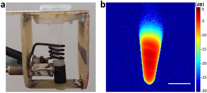
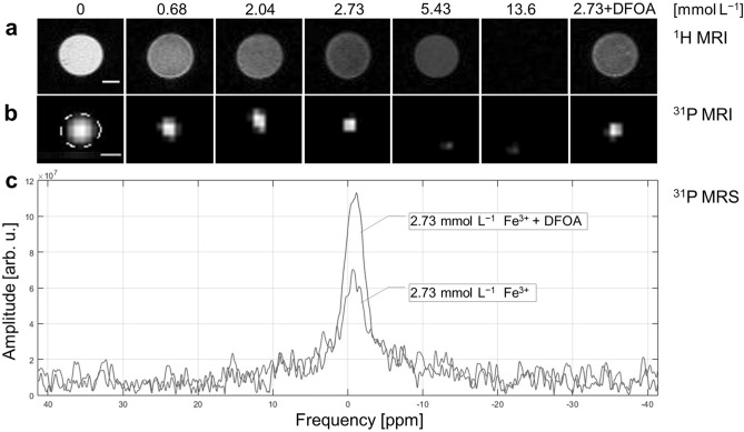
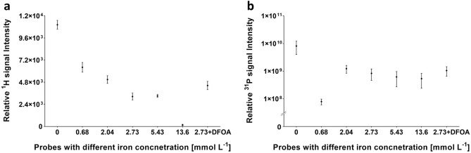
We present the MR properties of a novel bio-responsive phosphorus probe doped with iron for dual proton and phosphorus magnetic resonance imaging (1H/31P-MRI), which provide simultaneously complementary information. The probes consist of non-toxic biodegradable calcium phytate (CaIP6) nanoparticles doped with different amounts of cleavable paramagnetic Fe3+ ions. Phosphorus atoms in the phytate structure delivered an efficient 31P-MR signal, with iron ions altering MR contrast for both 1H and 31P-MR. The coordinated paramagnetic Fe3+ ions broadened the 31P-MR signal spectral line due to the short T2 relaxation time, resulting in more hypointense signal. However, when Fe3+ was decomplexed from the probe, relaxation times were prolonged. As a result of iron release, intensity of 1H-MR, as well as the 31P-MR signal increase. These 1H and 31P-MR dual signals triggered by iron decomplexation may have been attributable to biochemical changes in the environment with strong iron chelators, such as bacterial siderophore (deferoxamine). Analysing MR signal alternations as a proof-of-principle on a phantom at a 4.7 T magnetic field, we found that iron presence influenced 1H and 31P signals and signal recovery via iron chelation using deferoxamine.
© 2022. The Author(s).
Conflict of interest statement
The authors declare no competing interests.
Figures

Similar articles
-
Iron-based compounds coordinated with phospho-polymers as biocompatible probes for dual 31P/1H magnetic resonance imaging and spectroscopy.Sci Rep. 2024 Feb 15;14(1):3847. doi: 10.1038/s41598-024-54158-x. Sci Rep. 2024. PMID: 38360883 Free PMC article.
-
Iron(III)-Based Magnetic Resonance-Imageable Liposomal T1 Contrast Agent for Monitoring Temperature-Induced Image-Guided Drug Delivery.Invest Radiol. 2016 Nov;51(11):735-745. doi: 10.1097/RLI.0000000000000297. Invest Radiol. 2016. PMID: 27309776
-
Preliminary evaluation of iron phytate (inositol hexaphosphate) as a gastrointestinal MR contrast agent.J Magn Reson Imaging. 1993 Jan-Feb;3(1):119-24. doi: 10.1002/jmri.1880030120. J Magn Reson Imaging. 1993. PMID: 8428077
-
19F Magnetic Resonance Activity-Based Sensing Using Paramagnetic Metals.Acc Chem Res. 2020 Jan 21;53(1):2-10. doi: 10.1021/acs.accounts.9b00352. Epub 2019 Dec 6. Acc Chem Res. 2020. PMID: 31809009 Review.
-
The pharmacokinetics of the lymphotropic nanoparticle MRI contrast agent ferumoxtran-10.Cancer Biomark. 2009;5(2):69-73. doi: 10.3233/CBM-2009-0579. Cancer Biomark. 2009. PMID: 19414923 Review.
Cited by
-
Phosphorus-Containing Polymers as Sensitive Biocompatible Probes for 31P Magnetic Resonance.Molecules. 2023 Mar 2;28(5):2334. doi: 10.3390/molecules28052334. Molecules. 2023. PMID: 36903579 Free PMC article.
-
Long-term in vivo dissolution of thermo- and pH-responsive, 19F magnetic resonance-traceable and injectable polymer implants.Nanoscale Adv. 2024 Apr 8;6(12):3041-3051. doi: 10.1039/d4na00212a. eCollection 2024 Jun 11. Nanoscale Adv. 2024. PMID: 38868824 Free PMC article.
-
Cationic fluorinated micelles for cell labeling and 19F-MR imaging.Sci Rep. 2024 Sep 30;14(1):22613. doi: 10.1038/s41598-024-73511-8. Sci Rep. 2024. PMID: 39349687 Free PMC article.
-
Iron-based compounds coordinated with phospho-polymers as biocompatible probes for dual 31P/1H magnetic resonance imaging and spectroscopy.Sci Rep. 2024 Feb 15;14(1):3847. doi: 10.1038/s41598-024-54158-x. Sci Rep. 2024. PMID: 38360883 Free PMC article.
References
-
- An L, Cai Y, Tian Q, Lin J, Yang S. Ultrasensitive iron-based magnetic resonance contrast agent constructed with natural polyphenol tannic acid for tumor theranostics. Sci. China Mater. 2021;64:498–509. doi: 10.1007/s40843-020-1434-1. - DOI
-
- Sedlacek O, et al. Fluorinated water-soluble poly(2-oxazoline)s as highly sensitive 19F MRI contrast agents. Macromolecules. 2020;53:6387–6395. doi: 10.1021/acs.macromol.0c01228. - DOI
Publication types
LinkOut - more resources
Full Text Sources
Research Materials
