. 2022 Feb 8;5(1):115.
doi: 10.1038/s42003-022-03063-y.

An extended motif in the SARS-CoV-2 spike modulates binding and release of host coatomer in retrograde trafficking

Affiliations

An extended motif in the SARS-CoV-2 spike modulates binding and release of host coatomer in retrograde trafficking

Debajit Dey et al. Commun Biol. .

Abstract

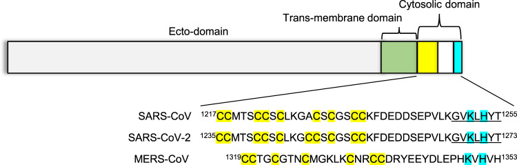
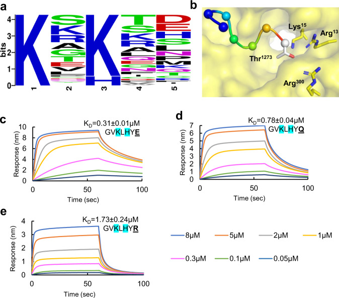
β-Coronaviruses such as SARS-CoV-2 hijack coatomer protein-I (COPI) for spike protein retrograde trafficking to the progeny assembly site in endoplasmic reticulum-Golgi intermediate compartment (ERGIC). However, limited residue-level details are available into how the spike interacts with COPI. Here we identify an extended COPI binding motif in the spike that encompasses the canonical K-x-H dibasic sequence. This motif demonstrates selectivity for αCOPI subunit. Guided by an in silico analysis of dibasic motifs in the human proteome, we employ mutagenesis and binding assays to show that the spike motif terminal residues are critical modulators of complex dissociation, which is essential for spike release in ERGIC. αCOPI residues critical for spike motif binding are elucidated by mutagenesis and crystallography and found to be conserved in the zoonotic reservoirs, bats, pangolins, camels, and in humans. Collectively, our investigation on the spike motif identifies key COPI binding determinants with implications for retrograde trafficking.


Conflict of interest statement

The authors declare no competing interests.

Figures

Fig. 1
Fig. 1. Organization of the coronavirus spike protein.
The spike protein is divided into an ecto-domain (gray), a trans-membrane domain (green), and a cytosolic domain (yellow-white-cyan). The cytosolic domain includes a cysteine rich tract (yellow) and a dibasic motif for COPI interactions (cyan). This overall organization and the dibasic motif are conserved in the spike protein of SARS-CoV, SARS-CoV-2, and MERS-CoV, which have been implicated in wide-spread human disease. The underlined residues correspond to the peptide sequence utilized in this manuscript.
Fig. 2
Fig. 2. Direct binding interaction of sarbecovirus spike hepta-peptide with αCOPI-WD40.
a Structural conservation of αCOPI-WD40 domain determined in the present investigation (yellow) and a previous structure (magenta). Arrow highlights main chain differences between these two αCOPI-WD40 structures in Gly168-Ala188. bh BLI assay of N-biotinylated spike hepta-peptide with COPI-WD40 domain. One representative experiment of three is shown in panels (bh). Color code for concentrations is given at the bottom of the figure. The equilibrium KD is provided with each sensorgram for comparison. b The spike wild-type peptide sequence demonstrates dose-dependent binding to αCOPI-WD40 domain. c Scrambling of the hepta-peptide sequence abolishes binding suggesting sequence-specific interaction. d β’COPI-WD40 demonstrates no interaction with the immobilized hepta-peptide. The mutant peptide, Gly-Val-Lys-Leu-Lys-Tyr-Thr, shows dose-dependent binding to (e) αCOPI-WD40 but not (f) β’COPI-WD40. g Acidification enhances binding between the wild-type spike hepta-peptide and αCOPI-WD40 domain. h β’COPI-WD40 shows weakly enhanced binding to the spike hepta-peptide upon acidification. “n.d.” implies not determined for weak interactions.
Fig. 3
Fig. 3. Structure-guided mutagenesis of spike hepta-peptide and binding analysis with αCOPI-WD40 domain.
a In silico model of the spike hepta-peptide complexed with αCOPI-WD40 domain (yellow surface). The hepta-peptide is shown as a ribbon in rainbow colors from N (blue) to C (red) terminus. The Cα-atoms in the hepta-peptide are shown as spheres. The side chains of residues that interact with αCOPI-WD40 are shown as a stick. bg BLI analyses of αCOPI-WD40 binding to spike hepta-peptide mutants. The color code of BLI traces is given at the bottom of the figure. One representative experiment of three is shown. Color code for concentrations is given at the bottom of the figure. The mutation in the spike hepta-peptide sequence is highlighted in bold and is underlined. The equilibrium KD is provided with each sensorgram for comparison. Mutagenesis of, (b) Lys1269, (c) His1271, or (d) both abolishes binding to αCOPI-WD40. e In contrast, Tyr1272 → Ala mutation only weakens binding to αCOPI-WD40. The middle panel shows weak binding of αCOPI-WD40 domain with a hepta-peptide wherein Lys1269 has been mutated to Ala. f Mutagenesis of Thr1273 to Ala in the spike hepta-peptide leads to moderately enhanced binding to αCOPI-WD40 whereas mutagenesis to Val1273 weakens binding (g).
Fig. 4
Fig. 4. In silico and biophysical analysis of spike C-terminal position in αCOPI-WD40 binding.
a Sequence logo generated from the alignment of K-x-H(K)-x-x sequence in 119 proteins predicted to be in the human membrane proteome. This shows the abundance of Lys in the first and third positions, low frequency of aromatic residues in the penultimate position, and the abundance of Asp and Glu in the C-terminal position. b An in silico model of the spike hepta-peptide on αCOPI-WD40 (yellow surface) shows an abundance of basic residues in the vicinity of the terminal Thr1273 spike residue. Panels (ce) show results of a BLI analysis of binding between spike hepta-peptide mutants and αCOPI-WD40. The equilibrium KD is provided with each sensorgram for comparison. Color code for concentrations is given at the bottom of the figure. Stabilization of the spike hepta-peptide complexed with αCOPI-WD40 is observed when the terminal position contains either, (c) acidic Glu1273, or (d) neutral Gln1273 residue. e In contrast, basic Arg1273 in the spike hepta-peptide does not favor enhanced binding. These data show a role of this terminal hepta-peptide position in modulating tight binding to αCOPI-WD40. In (ce), one representative experiment of three is shown.
Fig. 5
Fig. 5. Phylogenetic analysis of coronavirus spike C-terminal sequence.
This phylogram was generated from the alignment of five C-terminal residues in the spike protein. This penta-residue spike sequence is shown in italics to the right of each coronavirus. The residues in the dibasic motif are underlined. An acidic Asp is seen at the C-terminus of the spike proteins of only those coronaviruses that lack a dibasic motif.
Fig. 6
Fig. 6. Structure-guided mutagenesis of αCOPI-WD40 domain and binding analysis with spike hepta-peptide.
Panels (ac) highlight the hepta-peptide interactions (within 4 Å) of (a) Arg57, (b) Asp115, and (c) Tyr139 residues in αCOPI-WD40 in an in silico model. These three interacting αCOPI-WD40 residues are shown as yellow-red-blue sticks whereas the other residues are shown as a yellow surface for simplicity. The corresponding interacting residues in the spike hepta-peptide are labelled and shown as green-red-blue sticks and spheres for Cα atoms. The BLI analysis of Arg57 → Ala, Asp115 → Ala, and Tyr139 → Ala mutants with the wild-type spike hepta-peptide is shown in panels (d), (e), and (f), respectively. All three mutants demonstrate no substantial binding of the spike hepta-peptide. One representative experiment of three is shown in panels (df).

References

    1. Stadler K, et al. SARS-beginning to understand a new virus. Nat. Rev. Microbiol. 2003;1:209–218. - PMC - PubMed
    1. Ahmed AE. The predictors of 3- and 30-day mortality in 660 MERS-CoV patients. BMC Infect. Dis. 2017;17:615. - PMC - PubMed
    1. Zaki AM, van Boheemen S, Bestebroer TM, Osterhaus AD, Fouchier RA. Isolation of a novel coronavirus from a man with pneumonia in Saudi Arabia. N. Engl. J. Med. 2012;367:1814–1820. - PubMed
    1. Polack FP, et al. Safety and efficacy of the BNT162b2 mRNA COVID-19 vaccine. N. Engl. J. Med. 2020;383:2603–2615. - PMC - PubMed
    1. USFDA. U.S. Food and Drug Administration. Moderna COVID-19 vaccine [FDA briefing document]. Silver Spring, MD: U.S. Food and Drug Administration, Vaccines and Related Biological Products Advisory Committee; 2020, https://www.fda.gov/media/144434/download (2020).

Publication types

MeSH terms