Review
. 2022 May;18(5):290-308.
doi: 10.1038/s41574-021-00629-4. Epub 2022 Feb 8.

Uterine bleeding: how understanding endometrial physiology underpins menstrual health

Affiliations
Review

Uterine bleeding: how understanding endometrial physiology underpins menstrual health

Varsha Jain et al. Nat Rev Endocrinol. 2022 May.

Abstract

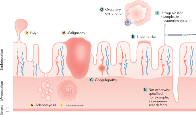
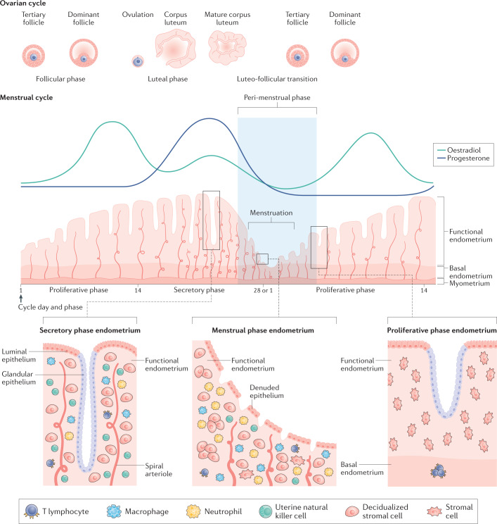
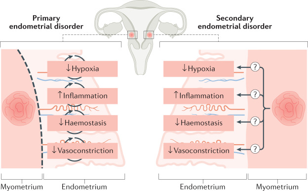
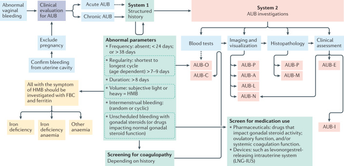
Menstruation is a physiological process that is typically uncomplicated. However, up to one third of women globally will be affected by abnormal uterine bleeding (AUB) at some point in their reproductive years. Menstruation (that is, endometrial shedding) is a fine balance between proliferation, decidualization, inflammation, hypoxia, apoptosis, haemostasis, vasoconstriction and, finally, repair and regeneration. An imbalance in any one of these processes can lead to the abnormal endometrial phenotype of AUB. Poor menstrual health has a negative impact on a person's physical, mental, social, emotional and financial well-being. On a global scale, iron deficiency and iron deficiency anaemia are closely linked with AUB, and are often under-reported and under-recognized. The International Federation of Gynecology and Obstetrics have produced standardized terminology and a classification system for the causes of AUB. This standardization will facilitate future research endeavours, diagnosis and clinical management. In a field where no new medications have been developed for over 20 years, emerging technologies are paving the way for a deeper understanding of the biology of the endometrium in health and disease, as well as opening up novel diagnostic and management avenues.


Conflict of interest statement

H.O.D.C. has received clinical research support for laboratory consumables and staff from Bayer AG, and provides consultancy advice (but with no personal remuneration) to Bayer AG, PregLem SA, Gedeon Richter, Vifor Pharma UK Ltd, AbbVie Inc. and Myovant Sciences GmbH. H.O.D.C. receives royalties from UpToDate for an article on abnormal uterine bleeding. V.J. receives salary and research consumables support from Wellbeing of Women (WoW). R.R.C. has been supported as a clinical research fellow by Bayer AG. J.A.M. receives salary and research consumables support from The Wellcome Trust.

Figures

Fig. 1
Fig. 1. FIGO System 2: the PALM-COEIN classification system.
The causes of abnormal uterine bleeding are classified using the acronym PALM-COEIN, with each letter denoting a cause. The structural causes (denoted by yellow letters) are discrete entities and include polyp, adenomyosis, leiomyoma (uterine fibroids) and malignancy. The non-structural causes (denoted by green letters) are depicted for the illustration; however, they cannot be measured or imaged. They include coagulopathy, ovulatory dysfunction, endometrial, iatrogenic and not otherwise classified (for example, a caesarean scar defect),. Note that the figure is a schematic and is not to scale.
Fig. 2
Fig. 2. Menstrual cycle and ovarian cycle physiology with highlighted aspects of the structure of the endometrium.
The endometrium is a dynamic structure that adapts to the endocrine environment in a cyclic manner, on average every 28 days. It undergoes a process of proliferation during an oestradiol-dominant phase. Following ovulation, the corpus luteum secretes progesterone, leading to a progesterone-dominant stage during which the endometrium decidualizes. In the absence of pregnancy, after the demise of the corpus luteum and progesterone withdrawal, the endometrium is shed during menstruation. This process requires the remaining cells to repair and regenerate without injury or scarring so that the menstrual cycle can repeat. As a multicellular tissue, the endometrium is highly responsive to the endocrine environment. The lower third of the endometrium, adjacent to the myometrium, is known as the basal layer and the upper two thirds, including the luminal surface, as the functional layer. Under the influence of changing levels of oestradiol and progesterone, the cellular structure of the endometrium adapts. The main cellular components within the endometrium are the epithelial cells, stromal cells, vascular cells and a variety of innate immune cells. The numbers of immune cells vary according to the cycle stage (see Figure key). The functional layer is shed during the menstrual phase, leaving behind a denuded basal endometrium. The peri-menstrual phase (also known as the luteofollicular transition) is the time after which progesterone and oestradiol levels fall, menstruation occurs and the endometrium transitions from a secretory to a proliferative state,,. Note that the figure is a schematic and is not to scale.
Fig. 3
Fig. 3. Major biosynthetic pathways in steroidogenesis.
The precursor of all steroid hormones is cholesterol. Steroidogenesis (synthesis of steroid hormones) occurs predominantly in the adrenal glands and gonads (ovaries and testes); however, it also occurs in the placenta. The key enzymes are located in the mitochondria and the endoplasmic reticulum of the cell. The first step in steroidogenesis involves the conversion of cholesterol to pregnenolone (a non-hormonal intermediate) within the cell mitochondria by the cholesterol side chain cleavage (CSCC) enzyme. The major classes of steroid hormones include progestogens, androgens, oestrogens and corticosteroids (glucocorticoids and mineralocorticoids). All steroid hormones have a cyclopentanoperhydrophenanthrene ring, and are further classified based on the number of carbon atoms. Pregnanes have 21 carbon atoms and are called C21 steroids (progestogens and corticosteroids). Oestranes have 18 carbon atoms (C18 steroids) and include oestrogens. Androstanes have 19 carbon atoms (C19 steroids) and include androgens. OH, hydrolase; HSD, hydroxysteroid dehydrogenase; DHT, dihydrotestosterone; DHEA, dehydroepiandrosterone; DHEAS, dehydroepiandrosterone sulfate.
Fig. 4
Fig. 4. The impact of the presence of uterine fibroids (leiomyoma) or adenomyosis on endometrial bleeding.
In the presence of myometrial conditions, such as uterine fibroids, it is unknown whether endometrial disorders that lead to an impairment in normal menstruation should be considered to be primary endometrial disorders (disorders that are independent of the myometrial condition). Primary endometrial disorders can include: a reduced local hypoxia response with consequent interference with coordinated endometrial repair; an increased inflammatory response after progesterone withdrawal; a reduced haemostasis response due to an overactive endometrial fibrinolytic response; and a decrease in local vasoactive factors limiting vasoconstriction of the spiral arterioles, thereby contributing to excessive bleeding. An alternative, unproven, hypothesis is that the endometrium might behave in a manner consistent with a ‘secondary endometrial disorder’, in which changes occur in endometrial function as a consequence of the presence of a myometrial condition (for example, uterine fibroids or adenomyosis).
Fig. 5
Fig. 5. Diagnostic pathway for abnormal uterine bleeding, and iron deficiency and/or iron deficiency anaemia.
On presentation with abnormal vaginal bleeding, the patient requires an initial assessment for pregnancy and to determine that the bleeding is coming from within the uterus, to confirm abnormal uterine bleeding (AUB). Such bleeding might either be acute or chronic (or acute on chronic). The time frame in which acute AUB is investigated varies, as the initial clinical priority is to stem the acute haemorrhage. In those with chronic AUB, it is necessary to perform a thorough assessment to determine the cause or causes in order to direct management. These include understanding the precise AUB symptoms using the International Federation of Gynecology and Obstetrics (FIGO) AUB System 1 (green boxes) prior to undertaking the appropriate investigations to identify the cause or causes of AUB using FIGO AUB System 2 classification (red boxes). Imaging and visualization can occur with one or a combination of transvaginal ultrasonography, sonohysterography, MRI and hysteroscopy. Of note, blood tests for AUB-C would follow a positive screen for coagulopathy. AUB-O is primarily diagnosed on the basis of the structured history and blood tests are generally not necessary, but measurement of serum levels of progesterone might be useful in selected instances. AUB-E is a primary disorder of endometrial haemostasis. The clinician makes this diagnosis if there is no other explanation found on the clinical assessment including history, physical examination, appropriate blood tests and uterine imaging. HMB, heavy menstrual bleeding; FBC, full blood count.

Similar articles

Cited by

References

    1. National Collaborating Centre for Women’s and Children’s Health. Heavy menstrual bleeding (Ch. 3). https://www.nice.org.uk/guidance/ng88/evidence/full-guideline-pdf-478229... (2007).
    1. Munro MG, et al. The two FIGO systems for normal and abnormal uterine bleeding symptoms and classification of causes of abnormal uterine bleeding in the reproductive years: 2018 revisions. Int. J. Gynecol. Obstet. 2018;143:393–408. doi: 10.1002/ijgo.12666. - DOI - PubMed
    1. Royal College of Obstetricians and Gynaecologists. National heavy menstrual bleeding audit final Report (Ch. 1). https://www.rcog.org.uk/globalassets/documents/guidelines/research--audi... (2014).
    1. Schoep ME, Nieboer TE, van der Zanden M, Braat DDM, Nap AW. The impact of menstrual symptoms on everyday life: a survey among 42,879 women. Am. J. Obstet. Gynecol. 2019;220:569.e1–569.e7. doi: 10.1016/j.ajog.2019.02.048. - DOI - PubMed
    1. Eaton SB, et al. Women’s reproductive cancers in evolutionary context. Q. Rev. Biol. 1994;69:353–367. doi: 10.1086/418650. - DOI - PubMed

Publication types