Review
. 2022 Oct;20(10):579-592.
doi: 10.1038/s41579-022-00683-3. Epub 2022 Feb 8.

The biogeography of infection revisited

Affiliations
Review

The biogeography of infection revisited

Sheyda Azimi et al. Nat Rev Microbiol. 2022 Oct.

Abstract

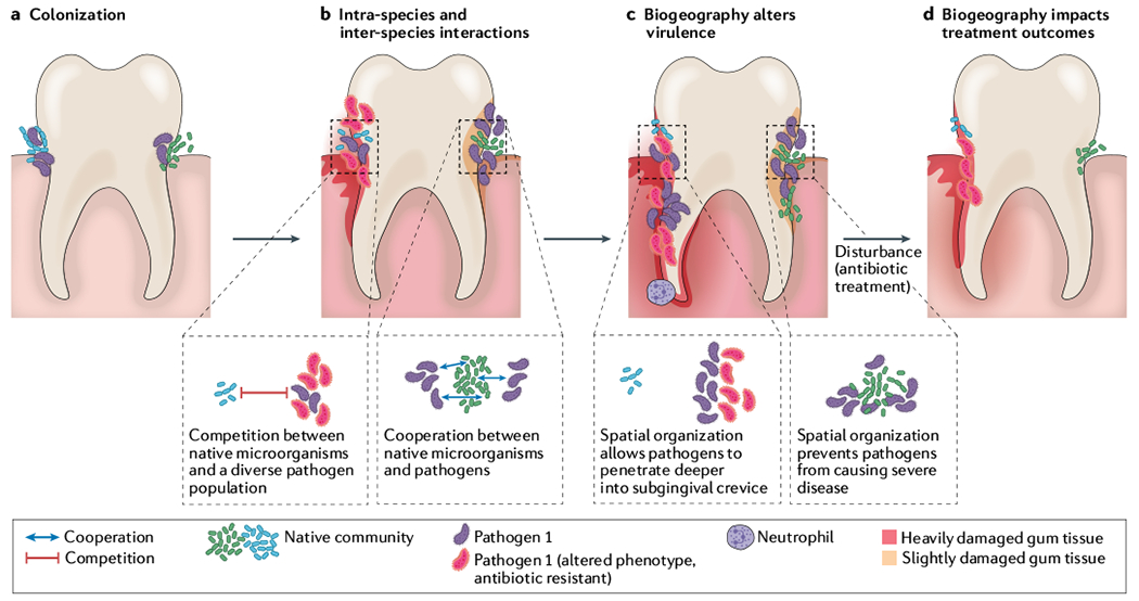
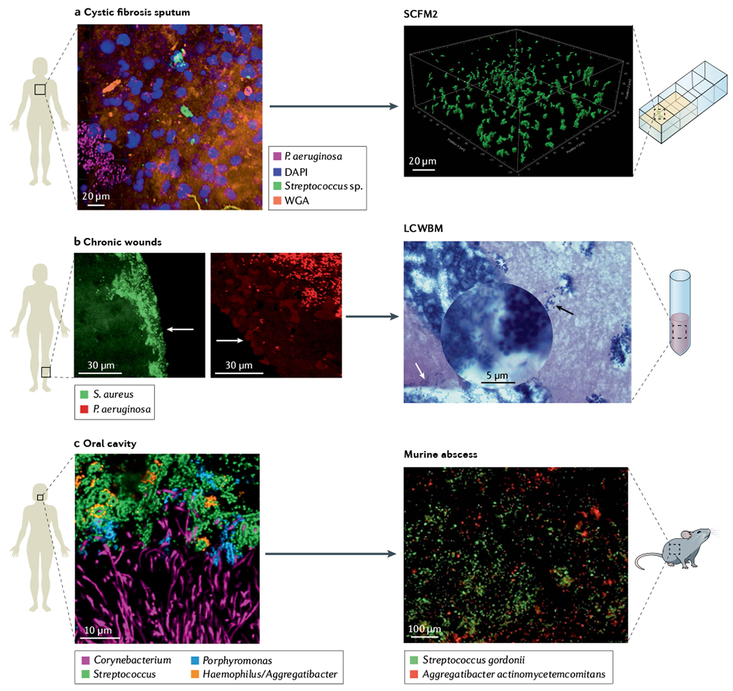
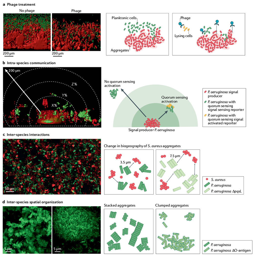
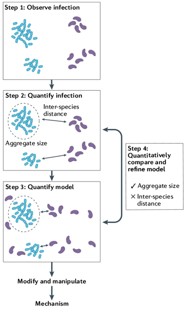
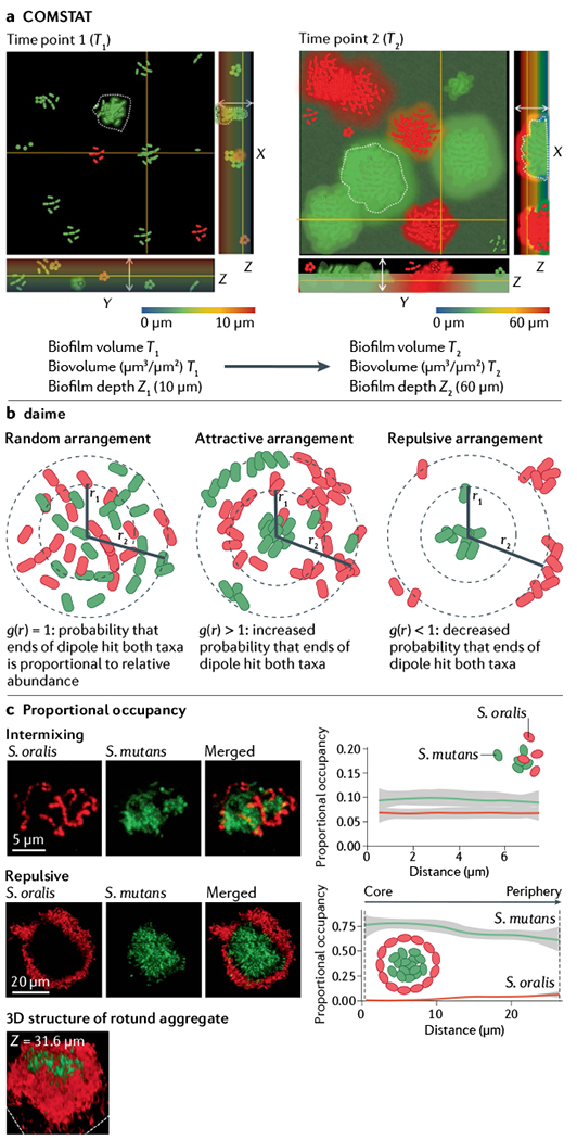
Many microbial communities, including those involved in chronic human infections, are patterned at the micron scale. In this Review, we summarize recent work that has defined the spatial arrangement of microorganisms in infection and begun to demonstrate how changes in spatial patterning correlate with disease. Advances in microscopy have refined our understanding of microbial micron-scale biogeography in samples from humans. These findings then serve as a benchmark for studying the role of spatial patterning in preclinical models, which provide experimental versatility to investigate the interplay between biogeography and pathogenesis. Experimentation using preclinical models has begun to show how spatial patterning influences the interactions between cells, their ability to coexist, their virulence and their recalcitrance to treatment. Future work to study the role of biogeography in infection and the functional biogeography of microorganisms will further refine our understanding of the interplay of spatial patterning, pathogen virulence and disease outcomes.


Conflict of interest statement

Competing interests

The authors declare no competing interests.

Figures

Fig. 1 |
Fig. 1 |. Polymicrobial biogeography can determine the severity of disease and treatment outcomes.
Spatial patterning of bacterial pathogens in relation to other microorganisms can alter the environment, microbial fitness and host immune responses. a | Colonization and aggregate formation by a pathogen (purple), along with the native microbial community (green and blue cells) in the subgingival pocket of the tooth. b | Expansion of the pathogen population can alter the environment by either limiting available nutrients or releasing exoproducts. Inter-species competitive (blue and purple cells) or cooperative (green and purple cells) dynamics determine infection biogeography. In addition, phenotypically and genetically diverse subpopulations can emerge (red cells); this heterogeneity can further alter the nature of intra-species interactions. c | Differential spatial patterning results in changes in virulence. Left side of tooth: localized expansion of pathogen leads to increased virulence, damaging the enamel (red enamel, root and gums) and resulting in periodontitis. This activity amplifies inflammatory signals and recruits immune cells such as neutrophils. In addition, increased pathogen abundance reduces available nutrients, decreasing fitness of the original microbial community. Right side of tooth: dispersed aggregates of the pathogen allow for increased fitness of the original microbial community, preventing both pathogen expansion and heavy damage to the subgingival pocket. d | Antimicrobial treatment is one of the most common disturbances for polymicrobial infections. Outcome of antimicrobial treatment is impacted by biogeography. On left side of tooth, spatial patterning protects the community from antimicrobial treatment. Further, the antibiotic increases fitness of an antibiotic-resistant pathogen (red cells), leading to niche expansion and increased damage. On right side of tooth, spatial patterning allows the pathogen to be susceptible to antibiotic treatment, eliminating the pathogen and reducing abundance of the original microbial community (green cells). This reduces virulence of the community and results in less damage to the tooth.
Fig. 2 |
Fig. 2 |. Microbiogeography of human-associated microbial communities is the benchmark for in vitro preclinical models.
a | Spatial arrangement of Pseudomonas aeruginosa (magenta) and streptococci (green) within a cystic fibrosis sputum sample visualized using MiPACT (microbial identification after passive clarity technique) and hybridization chain reaction. Structural context of the environment is visible using DAPI labelling of host DNA (blue) and wheat germ agglutinin (WGA) labelling of mucin (orange). Evaluation of cystic fibrosis sputum chemical composition and structure enable development of synthetic cystic fibrosis sputum medium (SCFM2), a medium that recapitulates the physical and chemical properties of cystic fibrosis sputum, where surface-detached aggregates of P. aeruginosa strain PAO1 in SCFM2 have similar spatial patterning as in cystic fibrosis sputum samples. b | P. aeruginosa (red) and Staphylococcus aureus (green) in two chronic wound biopsies visualized by peptide nucleic acid fluorescence in situ hybridization (PNA-FISH). Images show the spatial patterning of these two pathogens in wounds is not random. P. aeruginosa aggregates are localized deeper in wounds (50–60 μm from the surface) in comparison with S. aureus aggregates localized close to the wound surface (within 20–30 μm of the wound surface). Arrows show edge of wounds. Lubbock chronic wound biofilm model (LCWBM) provides a chemical environment similar to chronic wounds. However, spatial structure is dependent on microbial activities. Spatial arrangement of P. aeruginosa (white arrow) and S. aureus (black arrow) in this model is dependent on coagulase activity of S. aureus to turn soluble fibrinogen in plasma into insoluble fibrin, forming a fibrin network that gives gel-like properties to the media. In this model, S. aureus and P. aeruginosa can coexist in adjacent aggregates. c | Combinatorial labelling and spectral imaging FISH (CLASI-FISH) image of microbial communities from human supragingival plaque sampled from the human oral cavity showing a spatially patterned consortia of microorganisms in complex corncob structures. Investigations using a murine abscess model of infection demonstrated that precise spatial arrangement between Aggregatibacter actinomycetemcomitans and Streptococcus gordonii promotes virulence. Panel a cystic fibrosis sputum adapted from REF., CC BY 4.0 (https://creativecommons.org/licenses/by/4.0/). Panel b chronic wound adapted with permission from REF., ASM. Panel b LCWBM adapted with permission from REF., ASM. Panel c oral cavity adapted with permission from REF., PNAS. Panel c murine abscess adapted with permission from REF., PNAS.
Fig. 3 |
Fig. 3 |. Preclinical in vitro model SCFM2 provides a platform to study biological implications of microbiogeography.
Synthetic cystic fibrosis sputum medium (SCFM2) preclinical model of cystic fibrosis sputum has been a leader in linking biogeography with microbial interactions and fitness. a | Assessment of phage treatment efficacy showed that in SCFM2, planktonic migrant cells are sensitive to phage killing, whereas cells in aggregates and encased in exopolysaccharide matrix limit phage attachment and lysis. b | SCFM2 provides a structured environment to determine that a Pseudomonas aeruginosa aggregate of ~5,000 cells can signal to neighbouring aggregates up to 176 μm away, although the response to quorum sensing signalling can be heterogeneous,. In this experiment, only the aggregate in the bottom centre, confined in a micro-3D-printed trap, can produce the quorum sensing signal. When surrounding aggregates sense the quorum sensing signal, they express a fluorescent reporter gene (red in microscopy image, orange in schematic view). The quorum sensing signal is not detected in surrounding aggregates shown in green. c | Spatially structured environment of SCFM2 enables P. aeruginosa and Staphylococcus aureus aggregates to coexist, unlike in well-mixed environments. In this model, S. aureus aggregates are enriched at a distance of 3.5 μm from P. aeruginosa aggregates. S. aureus fitness did not change when co-cultured with the P. aeruginosa ΔpqsL mutant, which does not produce an anti-staphylococcal molecule. However, the average distance between S. aureus and P. aeruginosa ΔpqsL aggregates increased to 7.6 μm and antibiotic susceptibility of S. aureus increased, showing how changes in inter-species interactions and biogeography can impact S. aureus survival during infection. d | Loss of O-antigen is a common adaptation of P. aeruginosa to cystic fibrosis airways. This adaptation leads to changes in cell surface hydrophobicity, altering spatial patterning of cells in SCFM2. Cells with O-antigen are stacked, whereas those lacking O-antigen assemble into clumped aggregates, indicating that genetic variants of P. aeruginosa in cystic fibrosis airways can alter the biogeography of infection. Panel a adapted from REF., CC BY 4.0 (https://creativecommons.org/licenses/by/4.0/). Panel b reprinted from REF., CC BY 4.0 (https://creativecommons.org/licenses/by/4.0/). Panel c adapted from REF., CC BY 4.0 (https://creativecommons.org/licenses/by/4.0/). Panel d reprinted from REF., CC BY 4.0 (https://creativecommons.org/licenses/by/4.0/).
None
None

References

    1. Hall-Stoodley L, Costerton JW & Stoodley P Bacterial biofilms: from the natural environment to infectious diseases. Nat. Rev. Microbiol 2, 95–108 (2004). - PubMed
    1. Flemming HC & Wuertz S Bacteria and archaea on Earth and their abundance in biofilms. Nat. Rev. Microbiol 17, 247–260 (2019). - PubMed
    1. Azimi S, Klementiev AD, Whiteley M & Diggle SP Bacterial quorum sensing during infection. Annu. Rev. Microbiol 74, 201–219 (2020). - PubMed
    1. Ibberson CB & Whiteley M The social life of microbes in chronic infection. Curr. Opin. Microbiol 53, 44–50 (2020). - PMC - PubMed
    1. Martiny JB et al. Microbial biogeography: putting microorganisms on the map. Nat. Rev. Microbiol 4, 102–112 (2006). - PubMed

Publication types