

Study on Anatomical Variations in Fissures of Lung by CT Scan
- PMID: 35136490
- PMCID: PMC8817794
- DOI: 10.1055/s-0041-1741045
Study on Anatomical Variations in Fissures of Lung by CT Scan
Abstract
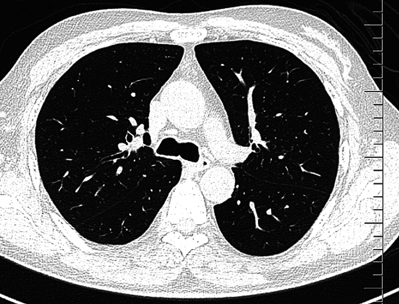
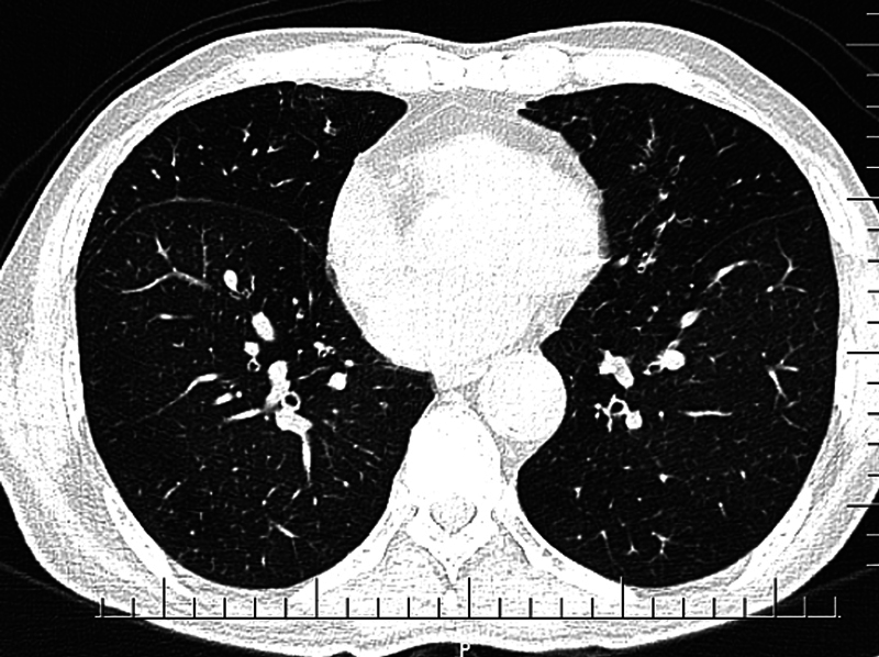
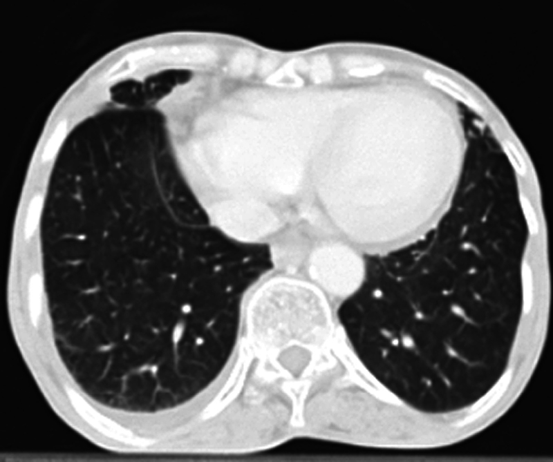
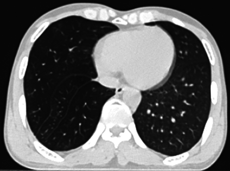
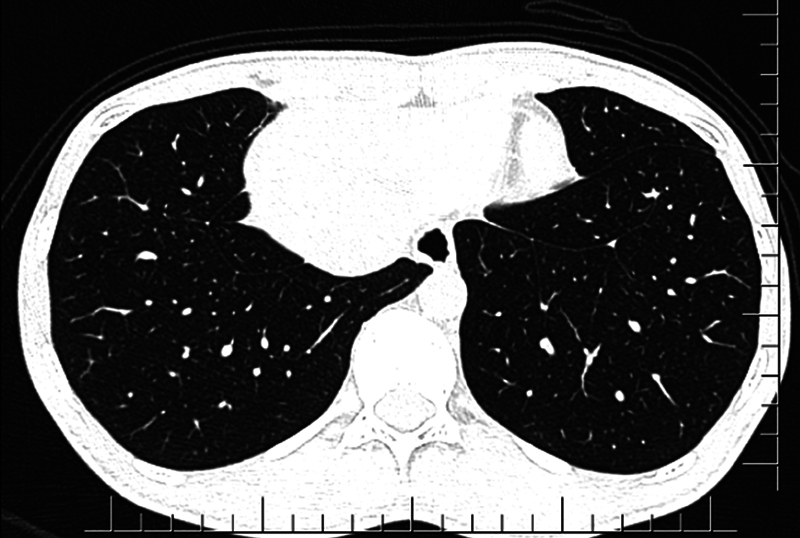
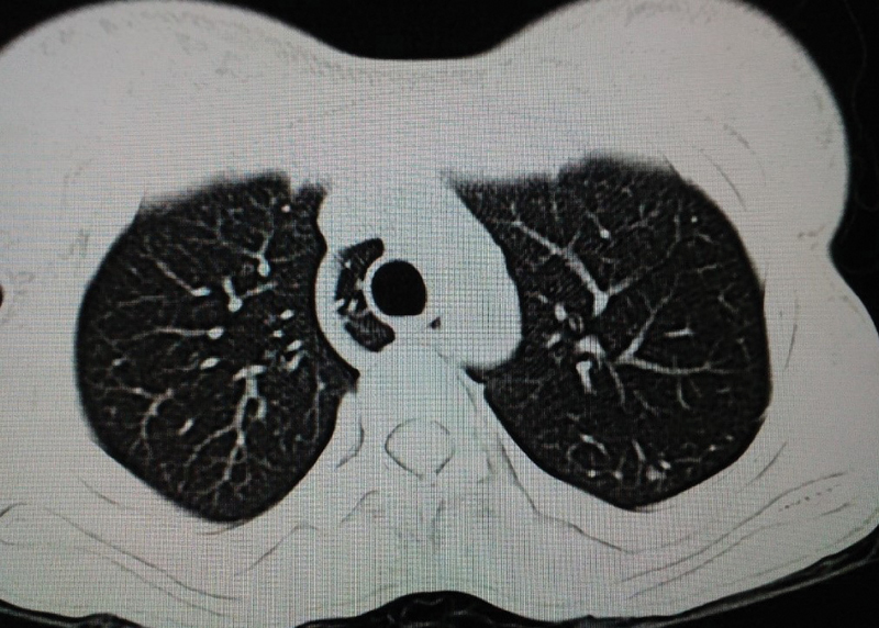
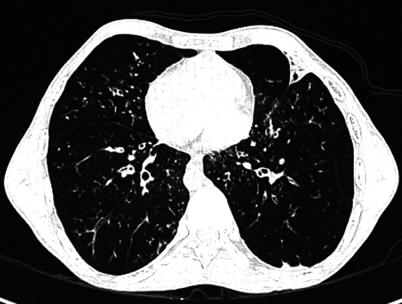
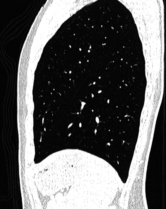
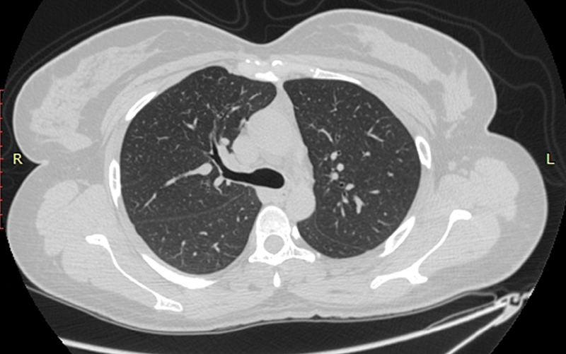
Introduction Refinements in the modern computed tomography (CT) imaging techniques have led to anatomical variations in the fissures of lung being diagnosed more frequently. So far, majority of the studies conducted are cadaveric. There is paucity of studies in this aspect based on chest CT images. Hence, we undertook this study to find the anatomical variations in the fissures. Prior detection of anatomical abnormalities is important to reduce postoperative complications in lung resection surgeries. Materials and Methods This was a cross-sectional study conducted over a period of 2 years. Data were collected from the patients who underwent CT scan thorax. Patients in whom normal anatomy of lung was distorted and cases where both lungs were not visualized completely were excluded from the study. All the CT images were reviewed by a single radiologist. The presence or absence of the normal and accessory pulmonary fissures, as well as the continuity of each fissure, was recorded by the radiologist. Data were compiled and analyzed. Results The study population consisted of 394 (70.4%) males and 166 (29.6%) females, totaling 560 cases. Fissural variations were detected in 22.9% ( n = 128). Also, 17.5% ( n = 98) fissural variations were seen in males and 5.4% ( n = 30) fissural variations were seen in females. Further, 54.7% ( n = 70) of variations were detected in the right lung and 45.3% ( n = 58) in the left lung. The most common fissural variation noted was right incomplete oblique fissure with a frequency of 8.4% cases ( n = 47). The most common accessory fissure detected was inferior accessory fissure. Total 22 cases were detected in both the lungs, 17 cases in male and 5 in female. Conclusion Anatomical variations in fissures were found to be more in the right lung than the left lung. Accessory fissures were detected in higher incidence on the right side.
Keywords: CT scan thorax; accessory fissures; anatomical variations; fissures of lung.
Indian Radiological Association. This is an open access article published by Thieme under the terms of the Creative Commons Attribution-NonDerivative-NonCommercial License, permitting copying and reproduction so long as the original work is given appropriate credit. Contents may not be used for commercial purposes, or adapted, remixed, transformed or built upon. ( https://creativecommons.org/licenses/by-nc-nd/4.0/ ).
Conflict of interest statement
Conflicts of Interest There are no conflicts of interest.
Figures

Similar articles
-
A pulmonary nodule mislocated in "dorsal" segment due to tri-lobed left lung.Front Surg. 2023 Jan 6;9:1069543. doi: 10.3389/fsurg.2022.1069543. eCollection 2022. Front Surg. 2023. PMID: 36684151 Free PMC article. Review.
-
Anatomical Variations in Pulmonary Fissures on Computed Tomography (CT).Cureus. 2022 Nov 30;14(11):e32062. doi: 10.7759/cureus.32062. eCollection 2022 Nov. Cureus. 2022. PMID: 36600863 Free PMC article.
-
Variations in pulmonary fissural anatomy: a medicolegal autopsy study of 256 cases.ANZ J Surg. 2020 Apr;90(4):608-611. doi: 10.1111/ans.15553. Epub 2019 Nov 10. ANZ J Surg. 2020. PMID: 31709740
-
Are textbook lungs really normal? A cadaveric study on the anatomical and clinical importance of variations in the major lung fissures, and the incomplete right horizontal fissure.Clin Anat. 2021 Apr;34(3):387-396. doi: 10.1002/ca.23661. Epub 2020 Aug 17. Clin Anat. 2021. PMID: 32713079
-
A comprehensive study and extensive review of morphological variations of liver with new insights.Surg Radiol Anat. 2022 Mar;44(3):455-466. doi: 10.1007/s00276-022-02883-1. Epub 2022 Jan 19. Surg Radiol Anat. 2022. PMID: 35048140 Review.
Cited by
-
A pulmonary nodule mislocated in "dorsal" segment due to tri-lobed left lung.Front Surg. 2023 Jan 6;9:1069543. doi: 10.3389/fsurg.2022.1069543. eCollection 2022. Front Surg. 2023. PMID: 36684151 Free PMC article. Review.
-
Morphological variations of fissures, lobes, and hilar pattern of the lung in a select South African sample.Surg Radiol Anat. 2024 Dec;46(12):2005-2017. doi: 10.1007/s00276-024-03497-5. Epub 2024 Oct 10. Surg Radiol Anat. 2024. PMID: 39387880 Free PMC article.
-
Anatomical Variations in Pulmonary Fissures on Computed Tomography (CT).Cureus. 2022 Nov 30;14(11):e32062. doi: 10.7759/cureus.32062. eCollection 2022 Nov. Cureus. 2022. PMID: 36600863 Free PMC article.
References
-
- Sadler T W. 11th ed. Baltimore, Maryland: Lippincott Williams & Wilkins; 2004. Langman's Medical Embryology; pp. 203–205.
-
- Magadum A, Dixit D, Bhimalli S. Fissures and lobes of lung – an anatomical study and its clinical significance. Int J Curr Res Rev. 2015;7:8–12.
-
- Tarver R D. How common are incomplete pulmonary fissures, and what is their clinical significance? AJR Am J Roentgenol. 1995;164(03):761. - PubMed
-
- Cronin P, Gross B H, Kelly A M, Patel S, Kazerooni E A, Carlos R C. Normal and accessory fissures of the lung: evaluation with contiguous volumetric thin-section multidetector CT. Eur J Radiol. 2010;75(02):e1–e8. - PubMed
-
- Craig S R, Walker W S. A proposed anatomical classification of the pulmonary fissures. J R Coll Surg Edinb. 1997;42(04):233–234. - PubMed
LinkOut - more resources
Full Text Sources
