. 2022 Jan 10;31(4):990-997.
doi: 10.1055/s-0041-1741046. eCollection 2021 Oct.

Case Series of Applications of Resting State Functional MRI in Brain Tumor Surgery: A Novel Technique

Affiliations

Case Series of Applications of Resting State Functional MRI in Brain Tumor Surgery: A Novel Technique

Arpita Sahu et al. Indian J Radiol Imaging. .

Abstract

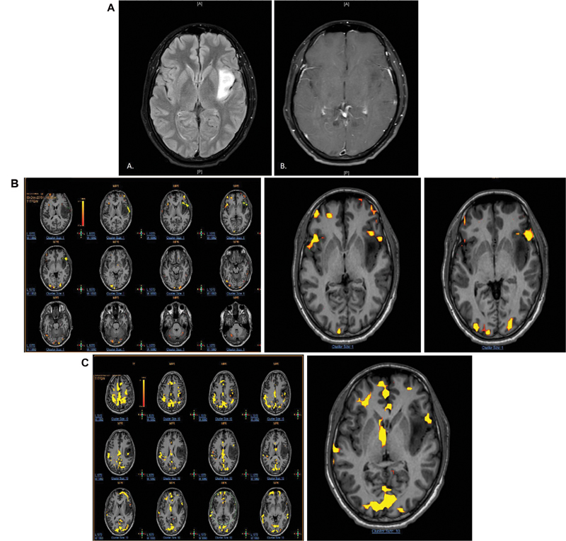
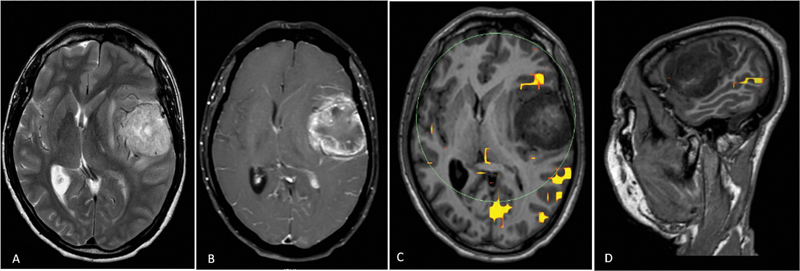
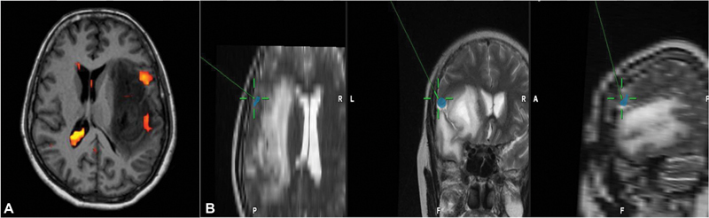
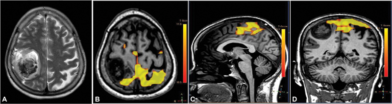
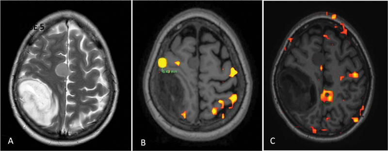
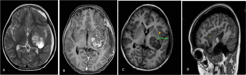
Background The extent of resection for brain tumors is a critical factor in determining the oncologic outcome for a patient. However, a balance between preservation of neurological function and maximal resection is essential for true benefit. Functional magnetic resonance imaging (fMRI) is one of the approaches that augments the neurosurgeon's ability to attain maximal safe resection by providing preoperative mapping. It may not be possible to perform awake craniotomy with intraoperative localization by direct cortical stimulation in all patients, such as children and those with neurocognitive impairment. Task-based fMRI may have limited value in these cases due to low patient cooperability. Methods In this article we present in a case-based format, the various clinical scenarios where resting state fMRI (rs-fMRI) can be helpful in guiding neurosurgical resection. rs-fMRI of the patients has been acquired on Philips 1.5 T system. Seed voxel method has been used for processing and analysis. Conclusion rs-fMRI does not require active patient cooperation to generate useful information and thus can be a promising tool in patients unable to cooperate for task-based studies.

Keywords: craniotomy; magnetic resonance imaging; resting state functional MRI.


Conflict of interest statement

Conflicts of Interest There are no conflicts of interest.

Figures

Fig. 1
Fig. 1
( A ) Axial T2-fluid attenuated inversion recovery image shows hyperintensity infiltrating glioma in the peri-insular region in the left frontotemporal lobe. ( B ) Axial postcontrast image through same section showing no contrast enhancement. ( C ) Task-based functional magnetic resonance imaging (fMRI) using word generation and verb generation paradigms was performed. Broca's area was seen on both the sides in the pars triangularis in the inferior frontal lobes suggestive of codominance. Broca's area on the left side is abutting the tumor on its anterior aspect. Resting state fMRI showing Broca's area only on the left side, abutting the tumor.
Fig. 2
Fig. 2
( A ) Axial T2 image showing hyperintense surface-based intra-axial lesion in the left frontotemporal region with T2 intermediate foci within. Perilesional edema and midline shift to the right are noted. ( B ) Axial T1 + C image showing thick peripheral shaggy rim enhancement of the lesion. ( C ) Resting state functional magnetic resonance imaging (rs-fMRI) superimposed on axial T1 images showing Broca's area placed anterior to the lesion. ( D ) rs-fMRI superimposed on sagittal T1 images showing Wernicke's area posteroinferior to the lesion.
Fig. 3
Fig. 3
( A ) Resting state functional magnetic resonance imaging (rs-fMRI) superimposed on axial T1-weighted images showing Broca's area in left pars triangularis, involved by the tumor. ( B ) Concordance between direct cortical stimulation (DCS) and rs-fMRI. Irregular blue region represents the Broca's area marked with rs-fMRI and green coordinates represent the area obtained by DCS. Images have been inverted for the convenience during surgery.
Fig. 4
Fig. 4
( A ) Axial T2 section showing heterogeneous right parietal lobe space occupying lesion. ( B ) Resting state functional magnetic resonance imaging (rs-fMRI) overlaid on axial T1-weighted image showing signal in the sensorimotor network. ( C and D ) rs-fMRI overlaid on coronal and sagittal T1-weighted images, respectively.
Fig. 5
Fig. 5
( A ) Axial T2-weighted image showing hyperintense lesion in right high parietal lobe with isointense right parafalcine meningioma. ( B ) Task-based functional magnetic resonance imaging (fMRI) superimposed on axial T1-weighted image showing 6 mm margin between tumor border and left hand representative area. However, there was no blood oxygen level dependent (BOLD) signal at expected supplementary motor area (SMA) region. ( c ) Resting state fMRI superimposed on axial T1-weighted image shows BOLD signal at SMA; the signal on contralesional side is more pronounced as compared with ipsilesional side due to plasticity.
Fig. 6
Fig. 6
( A ) Axial T2-weighted image showing iso- to hyperintense multilobulated lesion in left temporal lobe in a previously operated case showing subdural collection. ( B ) Axial T1-weighted postcontrast image showing patchy enhancement within the lesion. ( C and D ) Resting state functional magnetic resonance imaging superimposed, respectively, on axial and sagittal T1-weighted images showing Broca's area in the pars opercularis of the left inferior frontal lobe approximately at 1.4 cm anterior to the tumor.

Similar articles

Cited by

References

    1. Fisicaro R A, Jost E, Shaw K, Brennan N P, Peck K K, Holodny A I. Cortical plasticity in the setting of brain tumors. Top Magn Reson Imaging. 2016;25(01):25–30. - PMC - PubMed
    1. Bogomolny D L, Petrovich N M, Hou B L, Peck K K, Kim M J, Holodny A I. Functional MRI in the brain tumor patient. Top Magn Reson Imaging. 2004;15(05):325–335. - PubMed
    1. Jost E, Christiansen M H, Brennan N, Holodny A.Behavioral advantage in confrontation naming performance in brain tumor patients with left-frontal tumors In: Conference Proceedings of the Society for the Neurobiology of Language 2014;6:86
    1. Wood J M, Kundu B, Utter A et al.Impact of brain tumor location on morbidity and mortality: a retrospective functional MR imaging study. AJNR Am J Neuroradiol. 2011;32(08):1420–1425. - PMC - PubMed
    1. Biswal B, Yetkin F Z, Haughton V M, Hyde J S. Functional connectivity in the motor cortex of resting human brain using echo-planar MRI. Magn Reson Med. 1995;34(04):537–541. - PubMed