

Swiss cheese interventricular septum presenting with catastrophic stroke: the potential role of ECG-gated CTA
- PMID: 35136633
- PMCID: PMC8803220
- DOI: 10.1259/bjrcr.20210069
Swiss cheese interventricular septum presenting with catastrophic stroke: the potential role of ECG-gated CTA
Abstract
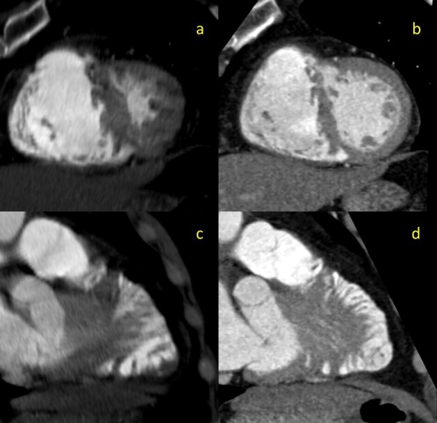
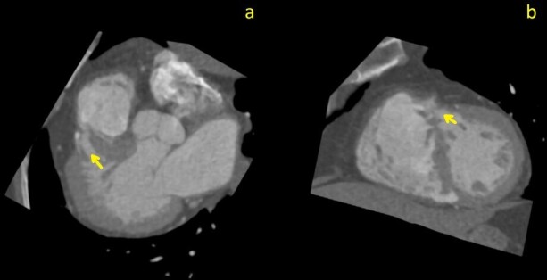
Ventricular septal defect is a common congenital cardiac condition that presents in a variety of morphologies. Less commonly, when an individual patient is found to have multiple ventricular septal defects, the term "Swiss cheese ventricular septal defect" is applied. Although not routinely utilized in clinical practice, electrocardiogram (ECG)-gated computed tomographic angiography (CTA) has been shown to provide utility in detecting intracardiac shunts, demonstrating promise in preventing acute strokes secondary to a paradoxical embolus from occurring; this is especially important when atypical cardiac septa are suspected. This case seeks to illustrate how usage of ECG-gated CTA can assist in early detection and prevention of adverse outcomes resulting from an atypical presentation of a ventricular septal defect.
© 2022 The Authors. Published by the British Institute of Radiology.
Conflict of interest statement
Competing interests: We wish to confirm that there are no known conflicts of interest associated with this publication.
Figures
Similar articles
-
Aneurysm of Mid and Apical Interventricular Cardiac Septum Dissecting Along the Basal Part - An Uncommon Entity Diagnosed with CT Angiography.Pol J Radiol. 2015 Oct 1;80:453-6. doi: 10.12659/PJR.895017. eCollection 2015. Pol J Radiol. 2015. PMID: 26516388 Free PMC article.
-
Swiss-cheese ventricular septal defect closure by combination sandwich patch.Asian Cardiovasc Thorac Ann. 2021 Jul;29(6):555-557. doi: 10.1177/0218492320976245. Epub 2020 Nov 24. Asian Cardiovasc Thorac Ann. 2021. PMID: 33231480
-
Surgical repair of multiple muscular ventricular septal defects: the role of re-endocardialization strategy.J Thorac Cardiovasc Surg. 2006 Nov;132(5):1072-80. doi: 10.1016/j.jtcvs.2006.07.011. J Thorac Cardiovasc Surg. 2006. PMID: 17059925
-
Cardiac septal aneurysm mimicking pseudomass: appearance on ECG-gated cardiac MRI and MDCT.AJR Am J Roentgenol. 2007 Jun;188(6):W550-3. doi: 10.2214/AJR.06.0996. AJR Am J Roentgenol. 2007. PMID: 17515346 Review.
-
Surgical management of muscular trabecular ventricular septal defects.Gen Thorac Cardiovasc Surg. 2011 Nov;59(11):723-9. doi: 10.1007/s11748-011-0826-9. Epub 2011 Nov 15. Gen Thorac Cardiovasc Surg. 2011. PMID: 22083689 Review.
References
-
- Mavroudis C, Dearani JA, Anderson RH. Ventricular septal defect. In: Atlas of Adult Congenital Heart Surgery: Springer, Cham; 2020. pp. 91–115.
-
- Minette MS, Sahn DJ. Ventricular septal defects [published correction appears in Circulation]. Circulation 2007;2006;114(20):2190–2197:618124. - PubMed
-
- Schwerzmann M, Windecker S, Meier B. Images in cardiovascular medicine. Swiss cheeselike atrial satrial septal defect. Circulation 1161;2008;117(24):e490–e492:757435. - PubMed
-
- Shuman WP, Leipsic JA, Busey JM, Green DE, Pipavath SN, Hague CJ, et al. . Prospectively ECG gated CT pulmonary angiography versus helical ungated CT pulmonary angiography: impact on cardiac related motion artifacts and patient radiation dose. Eur J Radiol 2012; 81: 2444–9. doi: 10.1016/j.ejrad.2011.06.017 - DOI - PubMed
Publication types
LinkOut - more resources
Full Text Sources
