Case Reports
. 2021 Aug 12;8(1):20210083.
doi: 10.1259/bjrcr.20210083. eCollection 2022 Jan 1.

Feasibility of arterial spin labeling in evaluating high- and low-flow peripheral vascular malformations: a case series

Affiliations
Case Reports

Feasibility of arterial spin labeling in evaluating high- and low-flow peripheral vascular malformations: a case series

Sanjeev Ramachandran et al. BJR Case Rep. .

Abstract

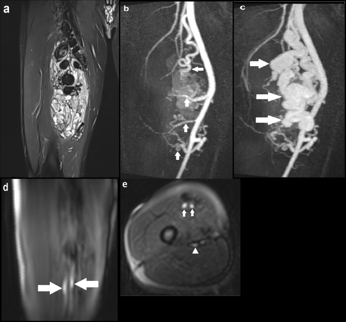
We present a case series highlighting a novel use of arterial spin labeling (ASL), a MRI perfusion technique, to evaluate both high- and low-flow peripheral vascular malformations (PVMs) across a range of anatomical locations. While the role of ASL in assessing intracranial vascular malformations is more established, there is limited evidence for PVMs. Our results provide preliminary evidence for the feasibility of ASL in imaging PVMs and its potential ability to distinguish between high- and low-flow PVMs. In addition, we demonstrate its ability to identify focal high blood flow, which may indicate the nidus in arteriovenous malformations. Together, these findings have important implications for patient management. We also outline the potential benefits and limitations of ASL in the imaging of PVMs, and provide justification for further validation of its diagnostic performance.


Conflict of interest statement

Competing interests: The authors declare that they have no conflict of interest.

Figures

Figure 1.
Figure 1.
a) Axial T2 image demonstrating a 18 × 16 mm hyperintense subcutaneous lesion in the left frontal scalp region, with multiple flow voids within. Underlying calvarium is intact; (b) axial MRA (TWIST) demonstrating robust early (arterial phase) enhancement of the lesion, with a draining vein shown at the same level (solid arrow). No communication with the intracranial circulation; (c) coronal MRA delineating an arterial feeder arising from the left superficial temporal artery (solid arrow); (d) axial ASL image demonstrating hyperintensity within the lesion, suggestive of high flow; (e) axial ASL image at a different slice delineating the draining vein (solid arrow), with the arterial feeder seen on MRA not identified. ASL, arterial spin labeling.
Figure 2.
Figure 2.
(a) Coronal STIR image demonstrating a large AVM with multiple large tortuous flow voids involving the vastus medialis and intermedius muscles; (b) coronal MIP image of dynamic MRA (TWIST) in the early arterial phase delineating four arterial feeders arising from the proximal, mid and distal SFA (solid arrows), with early enhancement of the AVM; (c) Subsequent MRA MIP image in the arterial phase demonstrating avid enhancement of the AVM with a large draining venous channel (solid arrows), multiple smaller venous channels and early filling of the distal SFV; (d) Coronal ASL image demonstrating two discrete foci of hyperintensity within the AVM (solid arrows), suggestive of the nidus through which blood flow is maximal; (e) Axial ASL image again highlighting the two foci of intense signal suggestive of the nidus (solid arrows). At the same level, there is another focus of less marked high signal posteriorly indicating early venous drainage (solid arrowhead); (f) Axial ASL image acquired at another slice identifying one of the superior arterial feeders (solid arrow). ASL, arterial spin labeling; AVM, arteriovenous malformation; MIP, maximum image projection; SFA, superficial artery; SFV, superficial vein; STIR, short tau inversion recovery.
Figure 3.
Figure 3.
(a) Catheter angiogram demonstrating an AVM involving the right hand with evidence of arteriovenous shunting; (b) catheter angiogram at a slightly later time point identifying two discrete niduses (solid arrows); (c, d) Axial ASL images at different slices demonstrating two separate foci of intensely high signal (solid arrows), corresponding with the niduses seen on catheter angiogram and providing evidence of high flow. ASL, arterial spin labeling; AVM, arteriovenous malformation.
Figure 4.
Figure 4.
(a) Axial STIR image demonstrating a 4.2 cm hyperintense lesion involving the right upper lip; (b) axial MRA (TWIST) image at approximately the same level showing maximal enhancement in the late images, with no definite arterial feeders or draining veins; (c) axial ASL image demonstrating normal signal intensity within the lesion. There is artefactual high signal at the outer border of the lip (not corresponding to the lesion) and elsewhere at the edges of the image; (d) carotid catheter angiogram showing maximal contrast opacification of the lesion in the delayed phase (solid arrow), with capillary type feeding vessels arising from the lingual artery and no large draining vein. No communication with the intracranial circulation. Appearances were in keeping with a low-flow verrucous vascular malformation, thought to be a predominantly venous malformation. ASL, arterial spin labeling; STIR, short tau inversion recovery.
Figure 5.
Figure 5.
(a) Axial T1 image demonstrating a small cluster of veins in the superficial subcutaneous tissue overlying the left gluteus maximus muscle (solid arrow), with no intramuscular component. There was fat suppression in between the cluster of veins (not shown); (b) axial T1 image at a different level showing one of the two draining veins traversing the gluteus maximum muscle (solid arrow), going onto communicate with normal-sized gluteal veins; (c) axial ASL demonstrating normal signal intensity in the region of interest (outline by a white circle). There is artefactual high signal at the edges of the image. ASL, arterial spin labeling.
Figure 6.
Figure 6.
(a) Axial STIR image demonstrating a small cluster of vessels in the subcutaneous tissue overlying the palmar aspect of the second metacarpal head, measuring 7 × 9 mm (solid arrow). No involvement of the underlying musculature; (b) coronal MIP image of dynamic MRA (TWIST) at the level of the lesion demonstrating no enhancement in the early arterial phase or arterial feeders (outline by the white circle); (c) axial ASL showing normal signal intensity in the region of interest (outline by the white circle). ASL, arterial spin labeling; MIP, maximum image projection; MRA, MR angiography; STIR, short tau inversion recovery.

Similar articles

References

    1. Hyodoh H, Hori M, Akiba H, Tamakawa M, Hyodoh K, Hareyama M. Peripheral vascular malformations: imaging, treatment approaches, and therapeutic issues. Radiographics 2005; 25 Suppl 1(Suppl 1): S159–71. doi: 10.1148/rg.25si055509 - DOI - PubMed
    1. Madani H, Farrant J, Chhaya N, Anwar I, Marmery H, Platts A, et al. . Peripheral limb vascular malformations: an update of appropriate imaging and treatment options of a challenging condition. Br J Radiol 2015; 88: 20140406. doi: 10.1259/bjr.20140406 - DOI - PMC - PubMed
    1. Deibler AR, Pollock JM, Kraft RA, Tan H, Burdette JH, Maldjian JA. Arterial spin-labeling in routine clinical practice, part 1: technique and artifacts. AJNR Am J Neuroradiol 2008; 29: 1228–34. doi: 10.3174/ajnr.A1030 - DOI - PMC - PubMed
    1. Detre JA, Alsop DC, Vives LR, Maccotta L, Teener JW, Raps EC. Noninvasive MRI evaluation of cerebral blood flow in cerebrovascular disease. Neurology 1998; 50: 633–41. doi: 10.1212/WNL.50.3.633 - DOI - PubMed
    1. Ramachandran S, Mukherjee D, Delf J, Bown MJ, Kandiyil N. A comparison of arterial spin labelling with catheter angiography in evaluating arteriovenous malformations: a systematic review. Br J Radiol 2020; 93: 20190830. doi: 10.1259/bjr.20190830 - DOI - PMC - PubMed

Publication types