

Case Reports
doi: 10.1259/bjrcr.20210085.
eCollection 2022 Jan 1.
Partial thrombosis of the corpus cavernosum - a malignancy mimicker
Affiliations
- PMID: 35136638
- PMCID: PMC8803217
- DOI: 10.1259/bjrcr.20210085
Item in Clipboard
Case Reports
Partial thrombosis of the corpus cavernosum - a malignancy mimicker
BJR Case Rep.
.
Abstract
Partial thrombosis of the corpus cavernosum is a rare condition, typically seen in young patients. Etiology, physiopathology and treatment are still not entirely understood. The authors report a case of a 49-year-old male with gastric cancer, who successfully treated a thrombosis of the corpus cavernosum conservatively. Diagnostic considerations and treatment options are discussed.
© 2022 The Authors. Published by the British Institute of Radiology.
Figures

Non-contrast CT (1a) shows an asymmetrical enlargement of the proximal segment of the right corpus cavernosum. In contrast-enhanced CT (1b), the mass remains hypodense with a hyperdense rim of enhancement, causing mass effect on the left corpus cavernosum.

Ultrasound (2a, 2c) at the base level of the penis revealed a well-circumscribed right-sided corpus cavernosum, enlarged and round-shaped, with heterogeneous decreased echogenicity. Doppler ultrasound (2b, 2d) showed no vascularity inside the abnormal image.

MRI (1.5 T) at the base of the penis. Axial view T1 weighted image shows the enlarged right corpus cavernosum, which is T1 hyperintense (*) comparing to the surrounding corpora. The left corpus cavernosum is displaced and compressed.
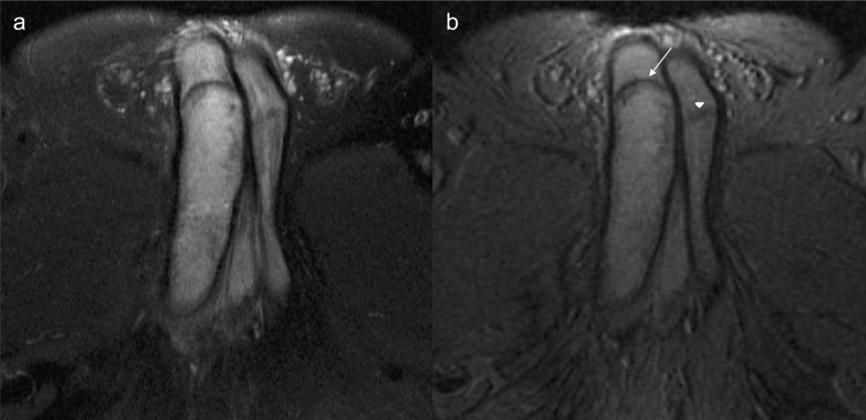
Axial T2 weighted FatSat (4a) and T2* images (4b) demonstrate iso/hyperintensity of the thrombus, due to extracellular methemoglobin. There is a membrane in the distal boundary of the thrombus displaying a low signal (arrow). There is a similar membrane in the contralateral corpus cavernosum (arrowhead).

Sagittal (5a) and coronal (5b) T1 weighted images with gadolinium contrast-enhanced demonstrate a lack of enhancement of the thrombosed right-sided corpus cavernosum, with a rim enhancement.
Similar articles
-
Bilateral partial thrombosis of the corpus cavernosum associated with the use of a stationary bike. Case report and literature review.Urol Case Rep. 2023 May 1;48:102414. doi: 10.1016/j.eucr.2023.102414. eCollection 2023 May. Urol Case Rep. 2023. PMID: 37215056 Free PMC article.
-
Partial segmental thrombosis of the corpus cavernosum.Cent European J Urol. 2011;64(4):264-5. doi: 10.5173/ceju.2011.04.art19. Epub 2011 Dec 9. Cent European J Urol. 2011. PMID: 24578910 Free PMC article.
-
A case of unprovoked segmental proximal partial thrombosis of the corpus cavernosum.Urol Case Rep. 2023 May 10;48:102426. doi: 10.1016/j.eucr.2023.102426. eCollection 2023 May. Urol Case Rep. 2023. PMID: 37215060 Free PMC article.
-
Idiopathic Partial Thrombosis (IPT) of the Corpus Cavernosum: A Hypothesis-Generating Case Series and Review of the Literature.J Sex Med. 2015 Nov;12(11):2118-25. doi: 10.1111/jsm.13036. Epub 2015 Nov 9. J Sex Med. 2015. PMID: 26553854 Review.
-
Partial segmental thrombosis of the corpus cavernosum: a case report and a review of the literature.Urology. 2012 Mar;79(3):708-12. doi: 10.1016/j.urology.2011.11.032. Urology. 2012. PMID: 22386425 Review.
Cited by
-
Utility of contrast enhanced ultrasound (CEUS) in penile trauma.Insights Imaging. 2023 Sep 25;14(1):158. doi: 10.1186/s13244-023-01499-2. Insights Imaging. 2023. PMID: 37749287 Free PMC article. Review.
References
Publication types
LinkOut - more resources
Full Text Sources
